Author Archives: Vicente Zaragozá

From Materials Research to Medical Manufacturing

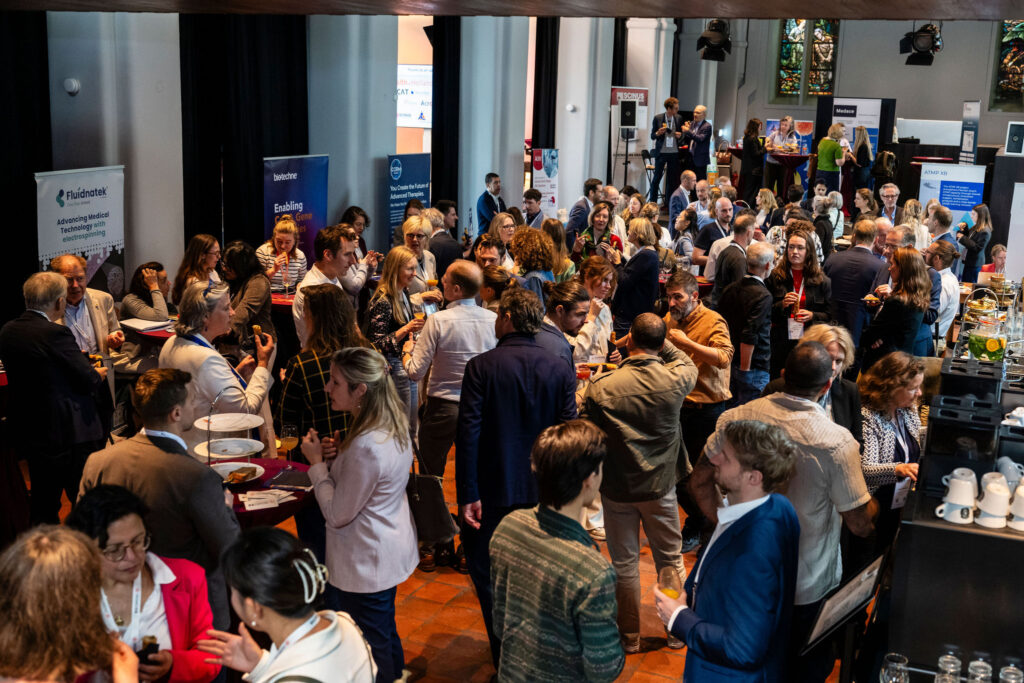
congress_Visitors

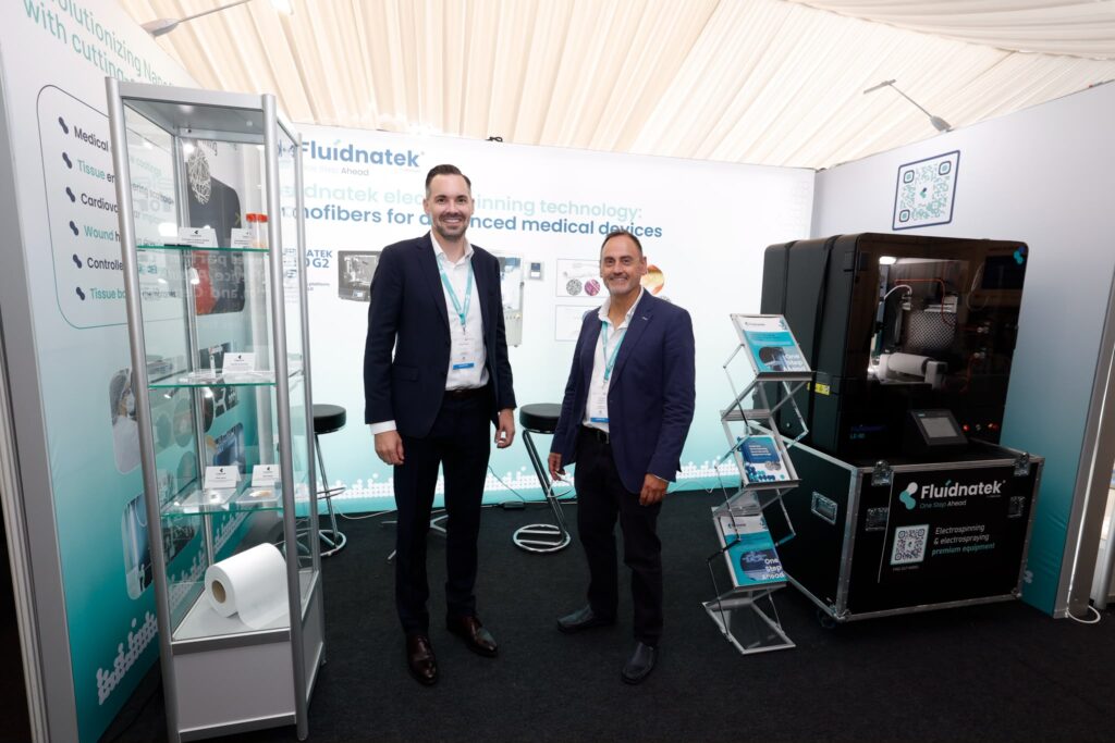
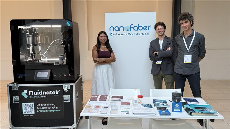
Innovation in electrospinning does not happen within a single discipline. It emerges through collaboration between materials scientists, biomedical researchers, medical device developers, and industrial manufacturers. Reflecting this multidisciplinary landscape, Fluidnatek will participate in five major European conferences throughout September, contributing to discussions that span the entire innovation journey—from materials research and process development to biomaterials, medical technologies, and industrial manufacturing

Dates and conferences:

  • Sept 1–4, 2026 — ELECTROSPIN (Padova, Italy)
    A specialist conference focused on electrospinning techniques, materials and applications. Ideal for researchers optimizing processes and for companies needing reproducible scaling and turnkey systems.
  • Sept 7–11, 2026 — 34th Annual Conference of the European Society for Biomaterials (Antwerp, Belgium)

A multidisciplinary biomaterials meeting covering preclinical testing to regulation and commercialization. A platform to engage with clinical groups and industry partners interested in implants and controlled drug delivery.

  • Sept 14–17, 2026 — XVIII Spanish Conference of Specialized Group on Polymers (GEP) (Valencia, Spain)

 Technical forum on polymers and applications, emphasizing innovation and tech transfer in Spain. An opportunity to showcase polymer formulations processable by electrospinning and discuss scaling with local R&D teams.

  • Sept 23–24, 2026 — MEDICAL TECHNOLOGY IRELAND (Galway, Ireland) Medical-technology event focused on industry–clinic collaboration and device supply chains. Perfect to present functional prototypes and manufacturing routes under regulatory constraints.
  • Sept 24–26, 2026 — GERMAN SOCIETY OF BIOMATERIALS (Aachen, Germany)

A rigorous biomaterials meeting with a strong orientation to clinical application and European standards. A key venue to validate electrospinning solutions with preclinical evaluators and manufacturing partners.

What we’ll showcase

  • Scalable, reproducible electrospinning equipment and processes, with performance data and traceability for regulated environments.
  • Live demonstrations of the Fluidnatek LE-50 Gen2 in operation, plus real collaboration case studies: from scaffolds for regeneration to drug-delivery patches and rapid prototyping tailored for industry needs.

We look forward to meeting researchers, manufacturers, and healthcare innovators throughout September to discuss their next electrospinning challenge. Whether the goal is process development, technology transfer, or industrial scale-up, our team will be available to explore practical solutions that help transform promising concepts into robust, reproducible manufacturing processes.

From Biomaterials to Medical Devices: Key Takeaways from Medical Technology Germany 2026

MedTechGermany_Fluidnatek

As medical technologies become increasingly sophisticated, manufacturing processes are evolving just as rapidly. Medical Technology Germany 2026 reflected this transformation, bringing together engineers, manufacturers, researchers, and regulatory specialists to discuss the technologies shaping the next generation of healthcare products.

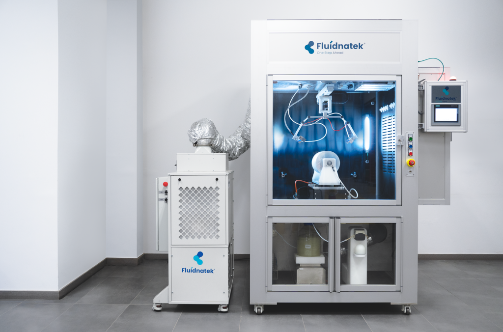
As part of this year’s exhibition, Fluidnatek participated in these discussions by showcasing the Fluidnatek LE-50 Gen2 electrospinning platform and engaging with professionals developing advanced medical devices, biomaterials, tissue engineering solutions, wound care products, drug delivery systems, and other healthcare technologies.

Electrospinning Continues to Gain Momentum

Many of the conversations held during the event reflected the growing interest in electrospinning as an enabling manufacturing technology for medical innovation.

By allowing precise control over nanofiber architecture, electrospinning enables the development of materials with highly tailored structural and functional properties. This level of control is becoming increasingly important for applications where porosity, surface area, mechanical performance, or controlled therapeutic release directly influence clinical outcomes.

As more medical products move from laboratory research toward commercialization, manufacturers are also placing greater emphasis on process reproducibility, scalability, and manufacturing robustness. These requirements extend beyond material design and increasingly influence technology selection from the earliest stages of product development.

From Research to Industrial Manufacturing

One of the recurring themes throughout Medical Technology Germany was the importance of establishing manufacturing strategies that can successfully transition from proof-of-concept studies to industrial production.

This transition was a recurring topic in many technical discussions throughout the event. While every project presents unique challenges, successful technology transfer often depends on combining robust process development with scalable manufacturing strategies.

These principles are reflected in many of the collaborative projects developed by Fluidnatek together with medical technology companies, research organisations, and industrial manufacturers, several of which are featured in our Customer Success Stories.

During the exhibition, the Fluidnatek team discussed how electrospinning projects can be developed with scale-up in mind from the outset. Rather than treating research and manufacturing as separate stages, an integrated development approach helps reduce technical risk, improve process understanding, and facilitate future technology transfer.

Visitors also had the opportunity to see the Fluidnatek LE-50 Gen2 operating live, demonstrating the level of process control, environmental stability, and automation required to achieve reproducible electrospinning results.

Supporting the Future of Medical Innovation

Medical technology continues to evolve rapidly, driven by increasingly sophisticated biomaterials, regenerative medicine, personalized therapies, and advanced drug delivery platforms. As these technologies mature, manufacturing processes must evolve alongside them to deliver the consistency, traceability, and quality required for successful commercialization.

At Fluidnatek, we remain committed to supporting researchers and manufacturers throughout this journey—from early feasibility studies and process development to scale-up and industrial manufacturing.

We would like to thank everyone who visited our booth during Medical Technology Germany 2026. We appreciate the opportunity to exchange ideas, discuss technical challenges, and explore how electrospinning can contribute to the next generation of medical technologies.

Fluidnatek at RegMed XB 2026: Driving the Transition from Lab to Clinical Reality

fluidnatek regmed

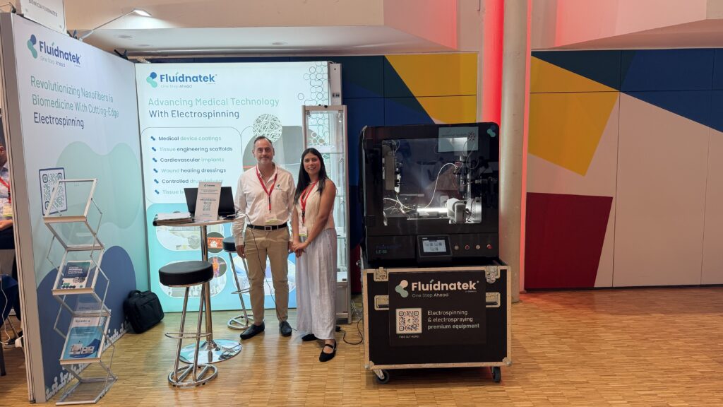
The RegMed XB Annual Conference 2026 in Utrecht has come to a close, and for Fluidnatek, it was much more than just a date on the calendar. Represented by our colleague Pilar Salinas, we spent several intensive days fully immersed in the heart of the regenerative medicine ecosystem, engaging with the pioneers who are redefining the future of healthcare.

Our presence at RegMed XB was driven by a clear purpose: to support the transition from groundbreaking research to real-world patient impact. 

The Translation Challenge

At Fluidnatek, we believe that the next generation of cell and gene therapies, tissue engineering, and advanced biomaterials depends entirely on the enabling technologies behind them. A brilliant scientific concept remains a concept until it can be manufactured under rigorous standards and moved into clinical applications.

During her interactions with researchers, clinicians, and ATMP developers, Pilar noted a common priority: bridging between proof-of-concept and clinical reality. Whether it’s developing complex scaffolds for tissue engineering or precision drug delivery systems, the consensus was clear—process control and scalability are the new frontiers.

Scaling Innovation with Precision

Our participation reinforced the vital role that electrospinning and electrospraying play in this ecosystem. These aren’t just laboratory techniques; they are scalable manufacturing platforms that offer:

  • Unmatched Precision: Creating nanofibrous structures that mimic the natural extracellular matrix.
  • Reproducibility: Essential for meeting the stringent requirements of regulatory bodies and clinical trials.
  • Scalability: Moving from benchtop research to pilot manufacturing without losing the properties that make the material effective.

Looking Ahead

Regenerative medicine is a collaborative journey. Seeing the entire value chain—from investors to policymakers and scientists—gathered in one place reminds us why we do what we do. At Fluidnatek, we are committed to providing the advanced processing technology that turns visionary research into tangible medical solutions.

The conference may be over, but the work of scaling the future of medicine continues. We are ready to help you navigate the manufacturing challenges of your next breakthrough.

Exhibitors regmed

Electrospun Membranes for Distillation

Electrospun nanofiber membrane structure used in membrane distillation

Introduction: The Challenge of Thermal Separation Processes

Water desalination, wastewater treatment, and resource recovery all depend on efficient separation technologies capable of producing high-purity water while minimizing energy consumption. Among the emerging approaches being investigated, membrane distillation has attracted significant interest because it combines high salt rejection with relatively low operating temperatures. This makes it a promising technology for advanced water treatment applications.

The efficiency of membrane distillation depends strongly on membrane structure and surface chemistry. Parameters such as porosity, pore size distribution, pore tortuosity, thickness, and hydrophobicity directly influence vapor transport and heat transfer resistance.

In recent years, electrospun membranes for membrane distillation have been extensively investigated because electrospinning enables the fabrication of highly porous nanofiber structures with interconnected pore networks. These architectures can facilitate vapor diffusion while maintaining separation between the feed and permeate streams.

As a result, electrospun nanofiber membrane distillation systems are becoming an increasingly important research area in advanced water treatment and separation science.

What Is Membrane Distillation?

Membrane distillation is a thermally driven membrane separation process in which a hydrophobic semipermeable membrane separates a heated feed solution from a colder permeate side.

The process operates through a vapor pressure gradient generated by a temperature difference across the membrane. Water evaporates at the interface between the warm feed and the membrane, and the vapors diffuse through the membrane pores and condense at the colder permeate side.

Because the membrane is hydrophobic, liquid water is prevented from penetrating the pores, resulting in selective vapor transport.

The performance of membrane distillation membranes depends on several key parameters:

  • Membrane porosity
  • Pore size distribution
  • Membrane thickness
  • Thermal conductivity
  • Hydrophobicity
  • Pore tortuosity

These factors influence vapor flux, conductive heat loss, liquid entry pressure (LEP; the maximum working pressure for the membrane distillation process, above which liquid water would penetrate the membrane), and long-term operational stability.

Traditionally, membrane distillation membranes are designed with relatively high thicknesses to reduce conductive heat transfer between the feed and permeate streams. However, thicker membranes may also increase the resistance to vapor transport, thus reducing the vapor flux.

Consequently, membrane engineering for distillation requires balancing thermal insulation with efficient vapor transport.

Electrospun Membranes for Membrane Distillation

Electrospinning is a versatile fabrication technology capable of producing continuous nanofibers from polymer solutions using a strong electric field.

Electrospun nanofiber membranes typically exhibit:

  • High surface-area-to-volume ratio
  • High porosity
  • Interconnected pore structures
  • Tunable fiber diameters

These structural characteristics are advantageous for membrane distillation because they can facilitate vapor diffusion.

At the same time, electrospun membranes must maintain sufficient hydrophobicity to prevent membrane wetting during operation.

Research has demonstrated that fiber morphology significantly influences membrane wetting behavior and vapor transport performance.

However, smoother fibers may sometimes provide higher vapor flux because of differences in pore interconnectivity and membrane packing density.

These observations highlight the importance of carefully balancing:

  • Fiber morphology
  • Hydrophobicity
  • Porosity
  • Mechanical stability
  • Liquid entry pressure

when designing electrospun membranes for distillation applications.

Hydrophobic Nanofibers for Efficient Vapor Transport

Hydrophobicity is one of the most critical parameters in membrane distillation systems. If pore wetting occurs (i.e., if liquid water penetrates the membrane pores), the feed and permeate solutions come in direct contact with each-other, resulting in contamination of the permeate stream. This scenario is considered a critical failure of the membrane.

Electrospun poly (vinylidene fluoride) (PVDF) membranes have been widely investigated in the context of membrane distillation due to their intrinsic hydrophobicity, chemical resistance, and thermal stability.

Experimental studies have shown that electrospun PVDF membranes can achieve relatively high vapor permeation fluxes. However, electrospun structures often exhibit lower liquid entry pressure than commercial PTFE membranes, meaning the risk of pore wetting is higher.

For example, reported studies observed that electrospun PVDF membranes exhibited liquid entry pressure (LEP) values below 0.64 bar, whereas commercial PTFE membranes demonstrated substantially higher LEP values of approximately 9 bar. The relatively low LEP of the electrospun PVDF membranes indicated a greater susceptibility to pore wetting under pressure-driven conditions, although these membranes often exhibited higher vapor flux due to their highly porous nanofibrous structure.

Despite this limitation, electrospun PVDF membranes have demonstrated higher vapor flux in certain membrane distillation configurations due to their highly porous nanofibrous architecture.

These findings indicate that electrospun nanofiber membranes may offer important mass transport advantages, although long-term wetting resistance remains an active area of research.

Materials and Design Strategies for High-Performance Membranes

Polymer Selection for Membrane Distillation

The polymer used during electrospinning strongly influences membrane performance in membrane distillation applications.

Materials commonly investigated include:

  • Polyvinylidene fluoride (PVDF)
  • Polyurethane (PU)
  • Polystyrene (PS)
  • Polysulfone (PSU)
  • PVDF-HFP copolymers

These polymers differ in:

  • Hydrophobicity
  • Thermal stability
  • Mechanical resistance
  • Solvent compatibility
  • Processability

Experimental studies with electrospun polyurethane (PU) membranes demonstrated that membrane thickness and feed salinity significantly influence permeation flux. In the work reported by Feng, Khayet, and Matsuura in their study on electrospun nanofibrous membranes for membrane distillation, thin electrospun membranes (approximately 6–10 g m⁻²) achieved fluxes above 10 kg m⁻² h⁻¹ under the experimental conditions reported by the authors and at feed salinities below 20 wt. % NaCl.

As feed salinity increased, flux progressively decreased to approximately 8 kg m⁻² h⁻¹, highlighting the impact of concentration polarization and membrane wetting phenomena on membrane distillation performance. In contrast, thicker electrospun membranes exhibited lower but more stable flux values, typically between 6 and 8 kg m⁻² h⁻¹ across the tested salinity range.

The lower stability of thinner membranes was associated with larger pore sizes and lower bubble-point pressures, increasing susceptibility to membrane wetting.

Surface Engineering and Hydrophobic Modification

Several surface engineering strategies have been investigated to improve hydrophobicity and wetting resistance in electrospun membranes.

One reported approach involved electroless silver plating followed by 1-dodecanethiol surface modification of electrospun PVDF nanofibers. The resulting membrane achieved:

  • Water contact angle ≈153°
  • Sliding angle <10°

Under the reported experimental conditions, the modified membrane maintained a relatively stable vapor flux during eight hours of membrane distillation testing.

Other studies (Zhou et al. (2014) have reported PTFE nanofibrous membranes fabricated by electrospinning PTFE/PVA precursor suspensions followed by high-temperature sintering. In this approach, PVA acts as a carrier polymer during electrospinning, while the PTFE particles are subsequently fused during thermal treatment to generate a nanofibrous PTFE structure.

These membranes demonstrated:

  • Water contact angle ≈156.7°
  • Salt rejection >98 %

during vacuum membrane distillation experiments.

Such approaches illustrate how membrane surface engineering can improve wetting resistance and operational stability in membrane distillation systems.


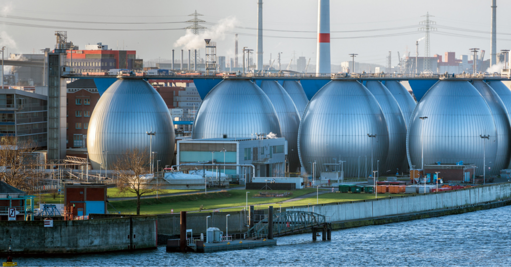
Desalination plant in Germany.

Desalination plant in Germany.

Performance and Efficiency Advantages

Nanofiber Membranes for Desalination and Water Treatment

Several experimental studies have evaluated electrospun membranes in desalination and water purification applications.

One investigation involving electrospun polysulfone nanofibers modified with beeswax reported:

  • Water contact angle ≈162°
  • Salt rejection >99.8 %
  • Permeate flux ≈6.4 L m⁻² h⁻¹

during direct contact membrane distillation testing with sodium chloride feed solution.

The membrane exhibited relatively stable operation over extended testing periods, with only moderate flux decline reported after prolonged operation.

These results demonstrate the potential of electrospun membranes for advanced water treatment applications where high salt rejection and controlled vapor transport are required.

Multi-Layer Membrane Architectures

To improve wetting resistance and liquid entry pressure, researchers have explored multilayer membrane designs.

One reported configuration included:

  • Hydrophilic nanofiber support layer
  • Cast membrane intermediate layer
  • Superhydrophobic electrospun PVDF top layer

In this structure:

  • The superhydrophobic layer reduces pore wetting
  • The intermediate layer increases liquid entry pressure
  • The hydrophilic support assists vapor transport

Under reported experimental conditions, the membrane maintained stable permeation performance over extended operating periods while preserving high salt rejection.

Influence of Fiber Architecture

Recent studies have also investigated electrospun membranes containing bimodal fiber diameter distributions.

These structures combine fibers of different diameters within the same membrane, modifying pore architecture and vapor transport pathways.

Research involving PS, PVDF-HFP, and blended systems reported permeation fluxes up to approximately 43.41 L m⁻² h⁻¹ while maintaining salt rejection near 99.74%.

These findings suggest that fiber diameter distribution and structural organization may significantly influence membrane distillation performance.

Fluidnatek’s Role in Electrospun Membrane Development and Scale-Up

Advanced electrospinning equipment plays an important role in the development of nanofiber membranes for membrane distillation and thermal separation technologies.

Fluidnatek electrospinning platforms support controlled fabrication of nanofiber membranes by enabling adjustment of:

  • Fiber diameter
  • Membrane thickness
  • Porosity
  • Polymer solution formulation
  • Multi-layer architecture

Such process control capabilities are relevant for researchers investigating membrane distillation, desalination membranes, hydrophobic nanofiber systems, and advanced filtration materials.

Fluidnatek platforms also support process scalability from laboratory development toward pilot-scale and even full-scale manufacturing configurations, which is an important consideration for membrane technology translation.

Conclusion

Membrane distillation is an important emerging technology for desalination and water treatment.

Electrospun membranes for membrane distillation offer several potential advantages, including:

  • High porosity
  • Tunable nanofiber structures
  • Enhanced vapor transport
  • Flexible membrane architecture design

At the same time, challenges related to pore wetting and long-term hydrophobic stability remain active areas of scientific research.

Ongoing developments in polymer engineering, multilayer membrane architecture, and surface modifications continue to improve the performance of electrospun nanofiber membranes for distillation applications.

Although many of these technologies are still at the research and pilot scale, electrospinning is expected to play an increasingly important role in next-generation membrane distillation systems.

Accelerate Membrane Distillation Research with Fluidnatek

Looking to develop high-performance membranes for distillation and advanced separation technologies?

Fluidnatek’s electrospinning platforms support the development of customizable nanofiber membranes for membrane distillation, desalination research, filtration, and other water treatment applications.

Contact Fluidnatek’s technical team to explore scalable electrospinning solutions for membrane engineering and thermal separation research.


References

ElectrospinTech. Electrospun membrane for distillation.
http://electrospintech.com/memdistillation.html

Essalhi, M., & Khayet, M. (2014). Surface modification of electrospun PVDF membranes for membrane distillation. Desalination.

Liao, Y., et al. (2013). Superhydrophobic modification of electrospun PVDF nanofibers for membrane distillation.

Zhou, X., et al. (2014). Electrospun PTFE nanofiber membranes for vacuum membrane distillation.

Prince, J. A., et al. (2014). Triple-layer membranes for improved membrane distillation performance.

Zhao, S., et al. (2023). Bimodal fiber diameter electrospun membranes for membrane distillation.

Khayet, M., & Matsuura, T. (2011). Membrane Distillation: Principles and Applications. Elsevier.

Feng, C., Khayet, M., & Matsuura, T. Preparation and Characterization of Electrospun Nanofibrous Membranes for Membrane Distillation. In: Membrane Distillation: Principles and Applications. Elsevier.


Electrospinning Environmental Control: How Temperature and Humidity Shape Fiber Morphology — and Why the ECU Makes the Difference

Fluidnatek ECU is critical for biomedical and GMP applications.

Environmental conditions strongly affect electrospinning outcomes, especially fiber diameter, morphology, and batch-to-batch reproducibility. Relative humidity and temperature influence solvent evaporation, jet stability, and fiber solidification, making environmental control a critical part of process development. Fluidnatek’s Environmental Control Unit (ECU) provides control of temperature and humidity to support more consistent electrospinning results across biomedical and research applications.

Introduction: Why Environmental Control Matters in Electrospinning

Electrospinning is highly sensitive to ambient conditions. Even when voltage, flow rate, and tip-to-collector distance are well optimized, changes in temperature and humidity can alter the way a jet forms and dries, which in turn affects final fiber morphology.

This sensitivity becomes especially important in applications where fiber uniformity matters, such as biomedical scaffolds, drug delivery systems, and protein-based biomaterials. In these cases, small changes in environmental conditions can lead to differences in fiber diameter, surface texture, and defect formation.

For this reason, environmental control is not simply a convenience. It is a practical tool for improving reproducibility, reducing process variability, and supporting more reliable electrospinning development. The Fluidnatek ECU is designed specifically for this purpose, giving users a controlled chamber environment that helps them work within a stable and adaptable climate window.

Mechanisms: How Humidity and Temperature Affect Fiber Morphology

Relative Humidity and Solvent Evaporation

Relative humidity plays an important role in electrospinning because it affects how quickly solvent evaporates from the jet and how the fiber surface solidifies. When humidity is too high, evaporation may slow down, which can increase the risk of surface pores, ribbon-like fibers, or other morphology changes. When humidity is too low, the jet may dry too quickly, which can also contribute to instability or defects in some systems.

The effect is not identical for every polymer or solvent system, but the broader principle is consistent: ambient humidity can significantly alter fiber formation. That is why controlled humidity is valuable when working with sensitive materials or when attempting to reproduce a specific morphology across multiple batches.

Temperature and Solution Properties

Temperature also influences electrospinning in several ways. It can affect solution viscosity, solvent vapor pressure, and the overall drying dynamics of the fiber jet. In many systems, a moderate increase in temperature can promote finer fiber formation by lowering viscosity and accelerating solvent evaporation.

At the same time, excessive heat can destabilize certain formulations, especially when working with protein-based or biologically sensitive materials. In those cases, process consistency depends not only on the electrospinning parameters themselves, but also on maintaining a stable and appropriate thermal environment throughout the run.


The Link Between Environment and Reproducibility

One of the biggest challenges in electrospinning is reproducibility. A process that works well on one day may behave differently on another if the lab environment changes. Seasonal shifts, air conditioning, ventilation, or simple humidity fluctuations can all influence the final fiber structure.

This is especially relevant when moving from exploratory research to more structured process development. If the goal is to compare formulations, optimize a scaffold, or build a scalable process, then environmental drift can make results harder to interpret. A controlled environment helps reduce this variability and makes it easier to isolate the effect of each process parameter.
The ECU addresses this need by providing active control over the chamber atmosphere. That gives researchers a more stable platform for comparing conditions, refining recipes, and documenting process behavior more consistently.


What the Fluidnatek ECU Brings to the Process

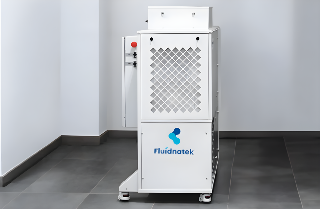
Fluidnatek’s Environmental Control Unit is designed to support electrospinning under controlled conditions by regulating temperature and humidity within the chamber. It is available as an integrated option across the Fluidnatek platform range and is intended to help users manage the climate conditions that affect fiber formation.
The ECU combines environmental stability with practical process support. According to Fluidnatek’s product positioning, it enables heating, cooling, drying, and humidifying of the chamber atmosphere, allowing users to explore the viable climate space for each process more effectively. This flexibility is particularly useful when working with polymers or solvents that respond differently to ambient conditions.

The system also supports a clean processing environment through HEPA filtration at the air intake stage. For biomedical and research applications, that added environmental control can be helpful when working toward more consistent and defensible results.


Applications Where Environmental Control Makes a Difference

Biomedical Scaffolds and Implantable Devices

In biomedical electrospinning, morphology matters. Fiber diameter, surface texture, and porosity all influence how a scaffold behaves in contact with cells and tissue. For applications such as wound dressings, vascular grafts, hernia meshes, and nerve guides, stable environmental conditions can help improve the consistency of the final material.

Polymer systems used in biomedical applications may be particularly sensitive to ambient changes. For that reason, the ability to control humidity and temperature during electrospinning can support more reliable scaffold development and more consistent batch-to-batch performance.


Drug Delivery and Functional Materials

Environmental control is also valuable in drug delivery work, where fiber morphology can affect loading behavior, surface characteristics, and release performance. In these cases, uncontrolled humidity may alter the way the fiber forms and dries, which can introduce unwanted variability.

Using a controlled chamber environment helps reduce one major source of uncertainty during process development. That makes it easier to compare formulations, evaluate design space, and make more confident decisions about process optimization.


Protein-Based Biomaterials

Protein-based electrospinning systems are often more sensitive than synthetic polymers. Materials such as collagen or gelatin may respond strongly to both temperature and humidity, which makes a stable processing environment even more important.

For these applications, environmental control can help preserve process consistency and reduce the likelihood of morphological defects. It also supports a more predictable workflow when researchers need to repeat experiments, compare formulations, or document results for publication or future scale-up.

Fluidnatek LE-500 + ECU_front view

Fluidnatek LE-500 and ECU.

Environmental Control and Scale-Up

Environmental control becomes even more important as electrospinning moves from lab-scale development to larger or more formalized production settings. At a larger scale, minor fluctuations in airflow, vapor accumulation, or chamber conditions can have a greater effect on fiber consistency.

A controlled chamber helps reduce one of the common variables that can complicate scale-up. By keeping temperature and humidity within a defined range, researchers and manufacturers can work toward more repeatable results across different systems and production settings.

This is where the ECU fits naturally into Fluidnatek’s broader platform approach. It is designed to support process development, product optimization, and the transition from exploratory research to more controlled production workflows.


Why the ECU Fits Fluidnatek’s Platform Strategy

The ECU is not a general-purpose climate accessory. It is part of a system designed specifically for electrospinning, where temperature, humidity, and airflow must be considered together.

That matters because electrospinning is not a static process. It is sensitive to the interaction between the solution, the jet, the collector, and the surrounding atmosphere. A controlled chamber gives users more flexibility to explore those interactions while reducing the noise introduced by uncontrolled ambient conditions.

For researchers, that means better experimental control and easier comparison between runs. For process developers, it means a more stable route toward reproducibility. And for teams working on biomedical or regulated applications, it helps support a cleaner and more consistent process environment.

Conclusion

Environmental control is a fundamental part of electrospinning process development. Temperature and humidity can strongly influence fiber morphology, diameter distribution, and reproducibility, making them essential variables to manage when working on high-value applications.

Fluidnatek’s Environmental Control Unit is designed to provide that stability within the electrospinning chamber. By supporting controlled heating, cooling, drying, humidification, and HEPA-filtered air intake, the ECU helps users create a more consistent processing environment for research and development.

For teams developing biomedical scaffolds, drug delivery systems, or other electrospun materials, that controlled environment can make the difference between a promising result and a repeatable process.

Looking to improve reproducibility in your electrospinning work? Fluidnatek’s Environmental Control Unit provides the controlled chamber conditions you need to support better fiber morphology, more consistent results, and more confident process development.

Contact our technical team to learn how the ECU can support your application.


References

Casper, C. L., Stephens, J. S., Tassi, N. G., Chase, D. B., & Rabolt, J. F. (2004). Controlling surface morphology of electrospun polystyrene fibers: Effect of humidity and molecular weight in the electrospinning process. Macromolecules, 37(2), 573–578. https://doi.org/10.1021/ma0351975

Nezarati, R. M., Eifert, M. B., & Cosgriff-Hernandez, E. (2013). Effects of humidity and solution viscosity on electrospun fiber morphology. Tissue Engineering Part C: Methods, 19(10), 810–819. https://doi.org/10.1089/ten.tec.2012.0671

Samad, U. A., Alam, M. A., Al-Zahrani, S. M., & Sherif, E. S. M. (2020). Effect of humidity on formation of electrospun polycaprolactone nanofiber embedded with curcumin using needleless electrospinning. Procedia Manufacturing, 43. https://doi.org/10.1016/j.promfg.2020.02.193

Xue, J., Wu, T., Dai, Y., & Xia, Y. (2019). Electrospinning and electrospun nanofibers: Methods, materials, and applications. Chemical Reviews, 119(8), 5298–5415. https://doi.org/10.1021/acs.chemrev.8b00593

Omer, S., Forgách, L., Zelkó, R., & Sebe, I. (2021). Scale-up of Electrospinning: Market Overview of Products and Devices for Pharmaceutical and Biomedical Purposes. Pharmaceutics, 13(2), 286. https://doi.org/10.3390/pharmaceutics13020286

Vass, P., et al. (2020). Scale-up of electrospinning technology: Applications in the pharmaceutical industry. WIREs Nanomedicine and Nanobiotechnology, 12(4), e1611. https://doi.org/10.1002/wnan.1611

Fluidnatek. (2024). Electrospinning Environmental Control Unit (ECU). Bioinicia Fluidnatek SLU. https://fluidnatek.com/advanced-electrospinning-equipment/electrospinning-environmental-control/

Fluidnatek, at the Spain–United States Business Summit

Boston biotech hub

Boston as a Global Biotech Innovation Ecosystem

Boston continues to position itself as one of the most concentrated and dynamic biotech hubs worldwide—bringing together leading research institutions, startups, established companies, and investors within a highly collaborative environment. This unique ecosystem makes the city a key reference point for advances in biomedical innovation, biotechnology, and medical device development.

At Fluidnatek, our participation in the Spain–United States Business Summit goes beyond event attendance. It is part of a broader strategy to remain closely aligned with the evolving needs of the biomedical and medical device sectors, particularly in areas where advanced materials and scalable manufacturing technologies are critical.

Strengthening Transatlantic Collaboration in Life Sciences

This initiative, promoted by ICEX in collaboration with Richi Foundation and local partners, brings together Spanish and US companies to foster collaboration in life sciences, biotechnology, and advanced technologies. Such platforms are essential to accelerate innovation and facilitate knowledge exchange across international ecosystems.

Boston_ICEX biotech summit

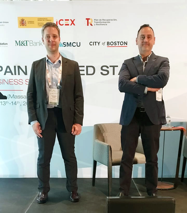
Fernando Corral -Operations Manager-, and Enrique Navarro -Sales & Marketing Manager, at the Spain-United States Business Summit.

Key Focus Areas in the Boston Life Science Ecosystem

In recent days, we have engaged with companies and innovation platforms across the Boston life sciences landscape—from early-stage ventures to well-established organisations—sharing perspectives on current challenges and opportunities in areas such as:

  • Advanced biomaterials and tissue engineering
  • Drug delivery systems and functional coatings
  • Scalable manufacturing of nanostructured materials

These domains are closely linked to the growing demand for electrospinning technologies and nanofiber-based solutions, particularly where performance, reproducibility, and scalability are key requirements.

Electrospinning and Nanofiber Technologies in Real-World Applications

Experiencing the level of innovation and commitment within the Boston ecosystem is truly inspiring. The energy surrounding these projects highlights the importance of translating research into practical, impactful solutions.

At Fluidnatek, the continuous improvement of our electrospinning platforms is directly shaped by these interactions. By listening to researchers and industry leaders, we ensure that our technology evolves in line with real application needs—from laboratory research to scalable nanofiber production.

Our presence in the US is well established. Over the years, we have supported this ecosystem by providing advanced nanofiber production technologies, enabling developments in biomedical applications, filtration, and beyond. Our commitment remains clear: to ensure that electrospinning moves beyond experimental settings and becomes a reliable, scalable manufacturing solution.

Driving Innovation Through Collaboration

Ultimately, meaningful innovation is driven by collaboration. Conversations with researchers, startups, and industry leaders are essential to identify challenges, refine technologies, and accelerate progress in the field of nanofiber-based biomedical applications.

👉 If you are working on biomedical applications involving nanofibers or electrospinning technologies, we invite you to get in touch with our team. Let’s explore how we can support your developments and collaborate on bringing scalable solutions to market.

Electrospun Tropoelastin Characterization: Fraunhofer IMWS Microscopy

electrospun tropoelastin characterization

Introduction: Characterizing Biomaterials at the Nanoscale

In regenerative medicine and biomedical engineering, the structural characteristics of biomaterials at the micro- and nanoscale play a decisive role in determining their biological performance. Cell adhesion, proliferation, and tissue integration are strongly influenced by surface morphology, mechanical properties, and scaffold architecture. Consequently, advanced biomaterial characterization techniques are essential for validating new materials intended for biomedical applications.

Among the many fabrication technologies used in tissue engineering, electrospinning has emerged as one of the most versatile methods for producing nanofiber scaffolds that resemble the fibrous architecture of the extracellular matrix (ECM). Electrospun biomaterials can provide highly porous, interconnected structures that support cell–material interactions and tissue regeneration (Almine et al., 2010).

To ensure that electrospun biomaterials meet the requirements for biomedical use, detailed microscopy analysis and nanofiber characterization are required. Techniques such as scanning electron microscopy (SEM) allow researchers to investigate fiber morphology, structural organization, and surface features at high resolution.

A collaborative research initiative involving Skinomics GmbH, Martin Luther University Halle-Wittenberg, and the Fraunhofer Institute for Microstructure of Materials and Systems IMWS is currently investigating innovative wound dressing materials based on human tropoelastin. The goal of this project is to develop customized biomaterials that combine biocompatibility, biodegradability, durability, and mechanical properties comparable to those of human skin (Fraunhofer IMWS, 2024). In this context, advanced Fraunhofer IMWS microscopy techniques are being used to characterize the structure and morphology of tropoelastin-based materials. Such characterization is a critical step in evaluating the biomedical potential of electrospun protein-based scaffolds.

Tropoelastin Nanofibers for Biomedical Applications and Tissue Engineering

What Is Tropoelastin and Why Is It Important in Biomedicine?

Tropoelastin is the soluble precursor of elastin, a structural protein that plays a fundamental role in maintaining the elasticity and resilience of many tissues in the human body. Elastin is present in the vasculature, skin, and lungs, where it enables tissues to stretch and return to their original shape (Wise, Mithieux & Weiss, 2009).

During biological processes, tropoelastin molecules assemble and crosslink to form elastin fibers. These fibers contribute to the mechanical stability and elasticity of tissues, making elastin an essential component of the extracellular matrix. Elastin is one of the most long-lived proteins in the human body, with a half-life that can span decades in tissues with low cellular turnover (Mithieux, Wise & Weiss, 2012).

According to the Fraunhofer IMWS project description, elastin is chemically and enzymatically stable, biocompatible, and inspires designs for tropoelastin-based materials with sustained mechanical performance in biological environments, given its long half-life in native tissues. These characteristics have motivated the development of tropoelastin-based biomaterials. By using the precursor protein of elastin, researchers aim to create materials that reproduce the mechanical and biological functions of native tissues (Fraunhofer IMWS, 2024).

As Dr. Christian Schmelzer, Head of the Department of Biological and Macromolecular Materials at Fraunhofer IMWS, stated: “Elastin is chemically and enzymatically extremely stable, biocompatible and does not produce immunological rejections when used as a biomaterial in humans. Therefore, we want to create new and innovative solutions for the treatment of complex wounds based on human tropoelastin” (Fraunhofer IMWS, 2024).

An additional motivation for developing tropoelastin-based materials lies in the limitations of conventional protein biomaterials derived from animal tissues. Animal-derived materials can pose risks of pathogen transmisión or unwanted immune reactions. Moreover, concerns about animal-derived medical products have increased among patients and healthcare providers. The use of human-derived recombinant tropoelastin may reduce such concerns while maintaining favorable mechanical and biological properties (Wise, Mithieux & Weiss, 2009).

Microstructure of a tropoelastin nonwoven

Microstructure of a tropoelastin nonwoven. Image: Fraunhofer IMWS

Electrospinning of Tropoelastin-Based Nanofibers

Electrospinning is widely used to fabricate electrospun biomaterials with nanoscale fibrous structures that mimic the architecture of the extracellular matrix. The technique involves the application of a high electric field to a polymer or protein solution, generating ultrafine fibers that are deposited as nonwoven membranes.

The electrospinning of tropoelastin has been studied as a method for producing porous protein scaffolds. Research has shown that the morphology of electrospun tropoelastin fibers can be modulated by varying spinning parameters such as delivery flow rate and starting protein concentration, and that cross-linked electrospun scaffolds retain the elasticity and cell-interactive properties inherent in the tropoelastin precursor (Wise, Mithieux & Weiss, 2009).

Electrospun scaffolds typically exhibit several characteristics that are advantageous for biomedical applications:

  • High surface-to-volume ratio
  • Porous fibrous architecture
  • Structural similarity to extracellular matrix networks
  • Tunable fiber diameter and scaffold morphology

In the Fraunhofer-associated research project, tropoelastin-based materials are being developed as customized wound dressing materials, specifically targeting the treatment of chronic and complex wounds. These conditions represent a major challenge in healthcare, particularly in aging populations. Chronic wounds such as venous ulcers, leg ulcers, and foot ulcers often require long-term treatment and can significantly affect patients’ quality of life (Fraunhofer IMWS, 2024). These conditions are also associated with considerable healthcare costs. Preclinical tests described in the Fraunhofer IMWS project documentation indicate that the tropoelastin-based material developed within the collaboration is a promising candidate for use as a wound dressing in such contexts. The material combines biocompatibility, biodegradability, durability, and favorable mechanical behavior.

Microscopy Techniques for Electrospun Biomaterial Characterization

SEM and Microscopy Techniques for Nanofiber Characterization Analyzing the structural properties of electrospun biomaterials requires detailed nanofiber characterization techniques capable of resolving features at the micro- and nanoscale. Microscopy methods are essential tools in biomaterials research for this purpose.

Among the techniques commonly used in the field to analyze electrospun scaffolds are:

  • Scanning Electron Microscopy (SEM): provides high-resolution images of fiber networks, enabling evaluation of fiber uniformity, diameter distribution, and structural integrity.

Transmission Electron Microscopy (TEM): allows investigation of internal fiber structure and nanoscale features.

  • Atomic Force Microscopy (AFM): used to assess surface topography and nanomechanical properties of individual fibers.
  • Optical microscopy: applied for preliminary morphological assessment and scaffold-level organization.

Scanning Electron Microscopy is instrumental analysis for evaluating electrospun protein-based scaffolds, as it enables high-resolution visualization of nanofiber topographies. Through SEM-based morphometric analysis, critical parameters—including fiber diameter distribution, uniformity, and overall architecture can be quantified, providing essential insights into the materials performance within biological environments.

Fraunhofer IMWS Research on Elastin-Like Materials

The Fraunhofer Institute for Microstructure of Materials and Systems IMWS specializes in the structural characterization of advanced materials. Within the tropoelastin research project, the institute applies microscopy techniques to analyze the morphology and structural properties of the developed biomaterials (Fraunhofer IMWS, 2024).

By combining electrospinning with detailed microstructural characterization, the research team can evaluate how the processing of tropoelastin-based materials influences their final structural and mechanical properties. These analyses provide insights into key aspects such as:

  • Fiber morphology and structural organization
  • Scaffold architecture and fiber distribution
  • Surface morphology of the nanofibers
  • Potential relationships between microstructure and material performance
Electrospun nonwoven of biotechnologically produced tropoelastin

Electrospun nonwoven of biotechnologically produced tropoelastin. Image: Fraunhofer IMWS.

Material Properties of the Tropoelastin-Based Wound Dressing

The collaborative project aims to create customized wound dressings based on tropoelastin biomaterials. According to the Fraunhofer IMWS project description, the developed material demonstrates a combination of properties that are particularly relevant for wound care applications (Fraunhofer IMWS, 2024):

  • Biocompatibility
  • Biodegradability
  • Mechanical properties comparable to human skin in preclinical assessments
  • Durability suitable for biomedical use

These characteristics are essential for wound dressing materials intended to support tissue regeneration while maintaining mechanical compatibility with the surrounding tissue. An important aspect highlighted in the project documentation is the similarity between the mechanical properties of the tropoelastin-based material and those of human skin—a property that can be attributed to the role of elastin in conferring skin elasticity and resilience (Wise, Mithieux & Weiss, 2009). Microscopy characterization is central to evaluating these structural properties. By analyzing nanofiber morphology and scaffold architecture, researchers can assess whether the material structure supports its intended biomedical function. The detailed characterization results from the Fraunhofer IMWS project are expected to be published in peer-reviewed literature as the project progresses.

Implications for Regenerative Medicine and Biomedical Device Development

The treatment of chronic and complex wounds represents a significant medical challenge, particularly in aging populations. Conditions such as venous ulcers and foot ulcers often require long-term care and can lead to serious complications if not treated effectively (Fraunhofer IMWS, 2024).

Innovative biomaterials are therefore being actively investigated to improve wound healing outcomes. Materials that combine biocompatibility, mechanical compatibility with surrounding tissues, and structural similarity to natural ECM are particularly promising. Tropoelastin-based biomaterials represent one such approach: because tropoelastin is the monomer of elastin, materials derived from it can reproduce structural and mechanical characteristics relevant to skin and other elastic tissues (Almine et al., 2010).

The use of human-derived recombinant protein materials also addresses concerns associated with animal-derived biomaterials, including potential infection risks and immune responses. This is a recognized advantage in the development of next-generation protein biomaterials for clinical use (Wise, Mithieux & Weiss, 2009).

The integration of electrospinning technologies with advanced biomaterial characterization enables researchers to systematically investigate these materials. Through high-resolution microscopy and structural analysis, researchers can evaluate whether electrospun tropoelastin scaffolds exhibit the morphological and mechanical properties required for biomedical applications.

Such interdisciplinary approaches—combining biomaterials science, nanofiber fabrication, and microscopy analysis—are central to the development of next-generation biomaterials for regenerative medicine and wound care.

Conclusion

The development of customized wound dressings based on human tropoelastin represents a significant and scientifically grounded direction in biomaterials research. By leveraging the properties of the soluble elastin precursor, researchers aim to create biomaterials that replicate the elasticity and resilience of natural skin tissue.

The collaborative project involving Skinomics GmbH, Martin Luther University Halle-Wittenberg, and Fraunhofer IMWS highlights the importance of combining biomaterial design with advanced structural characterization. Microscopy analysis

plays a key role in understanding the fiber morphology and scaffold architecture of electrospun tropoelastin materials, ensuring they meet the requirements for biomedical applications.

Continued advancements in the characterization and fabrication of tropoelastin-based materials may transform the treatment for non-healing wounds, offering a biomimetic foundation for innovative tissue engineering applications.

Looking to Develop and Validate Innovative Biomaterials Like Tropoelastin?

Fluidnatek supports advanced electrospinning projects for the production of electrospun biomaterials and nanofiber scaffolds tailored for biomedical research and regenerative medicine applications. Whether your project involves protein-based materials, ECM-mimicking scaffolds, or wound care devices, our platforms are designed to support rigorous research in collaboration with institutions such as Fraunhofer IMWS.

Contact Fluidnatek to discuss how our electrospinning solutions can support your biomaterial development pipeline.

References

Almine, J. F., Bax, D. V., Mithieux, S. M., Nivison-Smith, L., Rnjak, J., Waterhouse, A., Wise, S. G., & Weiss, A. S. (2010). Elastin-based materials. Chemical Society Reviews, 39(9), 3371–3379. https://doi.org/10.1039/b919452p

Fraunhofer Institute for Microstructure of Materials and Systems IMWS. (2024). Innovative wound care – customized wound dressings made from tropoelastin [Project communication]. Fraunhofer IMWS. https://www.imws.fraunhofer.de

Mithieux, S. M., Wise, S. G., & Weiss, A. S. (2012). Tropoelastin – a multifaceted naturally smart material. Advanced Drug Delivery Reviews, 65(4), 421–428. https://doi.org/10.1016/j.addr.2012.06.009

Wise, S. G., & Weiss, A. S. (2009). Tropoelastin. International Journal of Biochemistry & Cell Biology, 41(3), 494–497. https://doi.org/10.1016/j.biocel.2008.03.017

Wise, S. G., Mithieux, S. M., & Weiss, A. S. (2009). Engineered tropoelastin and elastin-based biomaterials. Advances in Protein Chemistry and Structural Biology, 78, 1–24. https://doi.org/10.1016/S1876-1623(08)78001-5

Blit, P. H., Battiston, K. G., Yang, M., Paul Santerre, J., & Woodhouse, K. A. (2012). Electrospun elastin-like polypeptide enriched polyurethanes and their interactions with vascular smooth muscle cells. Acta Biomaterialia, 8(7), 2493–2503. https://doi.org/10.1016/j.actbio.2012.03.032

Electrospun Fibers as Implant Interface Layer: Modulating Implant–Tissue Interactions

Implant–Tissue

Introduction: Enhancing Implant–Tissue Interactions

The long-term performance of biomedical implants is fundamentally determined by the biological response at the implant–tissue interface. Regardless of the bulk material used—metallic, polymeric, or composite—the surface in contact with host tissue governs protein adsorption, immune activation, cellular adhesion, and ultimately tissue remodeling. Suboptimal interface properties can result in persistent inflammation, fibrous encapsulation, or postoperative adhesions, compromising both functional outcomes and patient recovery.

In recent years, electrospun nanofiber membranes have emerged as promising candidates for engineering implant interface layers. Their structural resemblance to the extracellular matrix (ECM), combined with tunable surface chemistry and degradability, enables controlled modulation of cell–material interactions.

A recent study by Ren et al. (2023) investigated electrospun polycaprolactone (PCL)/polyethylene glycol (PEG) membranes as implant interface layers. By varying PEG content, the authors tailored membrane hydrophilicity and evaluated its influence on macrophage response in vitro and adhesion formation in vivo using a rat Achilles tendon injury model. Surface hydrophilicity emerged as a key factor in attenuating inflammatory signaling and optimizing tissue-implant integration

This article examines electrospun fibers as implant interface layers, focusing on their biological rationale, material strategies, translational relevance, and fabrication considerations within the biomedical context.

What Are Implant Interface Layers and Why Do They Matter?

An implant-tissue interface serves as a functionalized interlayer or biomimetic scaffold engineered to modulate the bidirectional biological signaling between a prosthetic device and surrounding tissue. Evolving beyond inert coatings, these architectures now function as bioactive modulators that influence the acute immune response and subsequent long-term homeostatic integration

Key biological processes occurring at the implant interface include:

  • Adsorption of serum proteins
  • Recruitment and activation of immune cells
  • Macrophage polarization dynamics
  • Fibroblast migration and extracellular matrix deposition
  • Fibrotic encapsulation or adhesion formation

Macrophages play a central role in determining the fate of implanted materials. Their polarization toward a pro-inflammatory (M1-like) or pro-regenerative (M2-like) phenotype significantly influences healing outcomes. Excessive or prolonged M1 activation is associated with chronic inflammation and fibrosis, whereas M2 polarization supports tissue repair and remodeling.

In the study by Ren et al., bone marrow-derived macrophages (BMDMs) cultured on electrospun PCL/PEG membranes exhibited hydrophilicity-dependent responses. Increasing PEG content enhanced membrane hydrophilicity and was associated with down-regulation of inflammatory gene expression and increased expression of markers linked to M2-like polarization. These results demonstrate that surface wettability can meaningfully influence immune cell behavior.

In vivo evaluation using a rat model further demonstrated that pure PCL membranes were associated with substantial adhesion formation, whereas PCL/PEG membranes showed reduced adhesion and facilitated easier separation of tendon from surrounding tissue. The membrane containing the highest PEG ratio exhibited the lowest inflammatory response and fewest adhesions among the tested groups.

Electrospun nanofibers are thus repositioned as bioactive transducers capable of governing tissue-to-implant integration, moving beyond the concept of static anatomical barriers

Electrospun Nanofibers for Implant–Tissue Integration

Electrospinning produces continuous fibers with diameters typically in the nano- to submicron range, forming porous, interconnected membranes. Several characteristics make electrospun nanofibers particularly attractive as implant interface layers.

Advantages of Fibrous Biointerfaces

  1. ECM-Mimetic Architecture

The fibrous morphology of electrospun membranes resembles native extracellular matrix, providing topographical cues that influence cell adhesion and morphology. This structural similarity can facilitate more physiological cell–material interactions compared to smooth or minimally textured surfaces.

  1. High Surface Area and Porosity

Electrospun mats present large surface areas for protein adsorption and cell contact, while their interconnected porosity supports nutrient diffusion and cellular infiltration where desired.

  1. Tunable Surface Chemistry

By blending polymers with different physicochemical properties, such as hydrophobic PCL and hydrophilic PEG, membrane wettability and degradation behavior can be adjusted. In the Ren et al. study, increasing PEG content directly modulated hydrophilicity and altered macrophage responses.

  1. Controlled Degradation

The study noted that membranes with higher PEG content exhibited a sparser multilayer structure in vivo, which may be related to faster degradation and potentially facilitated tissue separation at the membrane layer. This observation suggests that degradation kinetics can influence adhesion formation and interface remodeling.

Materials Used and Functionalization Strategies

H3 PCL/PEG Blended Systems

Polycaprolactone (PCL) is a widely used biodegradable polyester known for its mechanical flexibility and slow hydrolytic degradation.

Nevertheless, its inherent hydrophobicity frequently leads to non-specific protein adsorption, which may trigger adverse pro-inflammatory responses.

Polyethylene glycol (PEG), in contrast, is hydrophilic and widely used to enhance surface wettability and reduce non-specific protein adsorption. By blending PEG with PCL prior to electrospinning, Ren et al. created membranes with tunable hydrophilicity while maintaining structural integrity.

The study demonstrates that increasing PEG content:

  • Enhances hydrophilicity
  • Reduces inflammatory gene expression in macrophages
  • Promotes M2-like polarization
  • Reduces adhesion formation in vivo

Importantly, the investigation did not rely on additional bioactive molecules or drug incorporation; the modulation effect was achieved solely through adjustment of polymer composition and resulting surface properties.


Role of Fiber Alignment

The membranes in the study were described as aligned nanofibers. Fiber alignment can influence cell orientation and migration, particularly in musculoskeletal applications where anisotropic tissue architecture is critical. While the study focuses primarily on hydrophilicity effects, alignment may contribute to guiding tissue organization at the interface.

Considerations for Surface Modification

Beyond polymer blending, electrospinning platforms allow additional strategies such as incorporation of bioactive agents or post-fabrication surface treatments. However, the Ren et al. work specifically highlights that even without complex biochemical functionalization, physicochemical modulation alone can significantly alter immune response and adhesion outcomes.

Applications and Clinical Relevance

Tendon Repair and Adhesion Prevention

Postoperative adhesions remain a major complication in tendon surgery, limiting mobility and functional recovery. In the rat Achilles tendon injury model used by Ren et al., pure PCL membranes were associated with substantial tissue adhesion. In contrast, PCL/PEG membranes reduced adhesion formation, and the highest PEG ratio yielded the most favorable outcome in terms of reduced inflammation and improved tissue separation.

These findings suggest that electrospun implant interface layers may serve as physical and biological barriers that minimize pathological fibrotic bridging while supporting controlled healing.


Broader Implications for Implant–Tissue Integration

Although the study specifically evaluates a tendon model, the underlying principle—modulation of macrophage phenotype through surface hydrophilicity—has broader implications for other soft tissue implant applications. However, extrapolation to orthopaedic hard-tissue implants or cardiovascular devices requires dedicated experimental validation.

The work supports a paradigm in which implant surface engineering prioritizes immune modulation as a primary design objective.

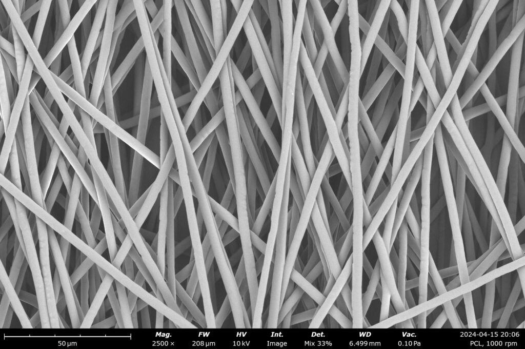
PCL aligned fibers made at 1000 rpm

PCL aligned fibers made at 1000 rpm. Image credit: Nanoscience Instruments.

Fluidnatek’s Capabilities in Implant Interface Nanofiber Development

Translating preclinical findings into practical biomedical applications requires reproducible fabrication platforms capable of controlling fiber morphology, alignment, and polymer composition.

Fluidnatek provides electrospinning systems designed to support:

  • Precise control of polymer blending (e.g., PCL/PEG ratios)
  • Fabrication of aligned nanofiber membranes
  • Reproducible control over fiber diameter and morphology
  • Development of degradable fibrous interface layers

Such platforms enable research teams to replicate and extend experimental configurations similar to those described by Ren et al., facilitating systematic studies on implant–tissue integration and immune modulation.

More information on electrospinning platforms for biomedical research is available at: https://fluidnatek.com/electrospinning-machines/

Conclusion: Toward Immunomodulatory Implant Interfaces

Electrospun fibers as implant interface layers represent a strategic evolution in biomedical surface engineering. Rather than functioning solely as passive structural coatings, these nanofiber membranes can actively influence early immune responses and subsequent tissue remodeling.

The study by Ren et al. demonstrates that tuning hydrophilicity through PCL/PEG blending modulates macrophage gene expression and reduces adhesion formation in a rat tendon injury model. Increased PEG content correlated with reduced inflammatory signaling, enhanced M2-like polarization, and fewer postoperative adhesions. Additionally, higher PEG ratios were associated with structural changes consistent with faster degradation, which may facilitate tissue separation at the interface.

These findings reinforce the concept that surface chemistry and nanoscale architecture are central determinants of implant performance. Continued investigation into electrospun nanofiber interface layers may advance the development of next-generation biomedical implants designed not only for mechanical function, but also for precise biological integration.

References

Ren, Y., et al. (2023). Electrospun fibers as implant interface layer. ElectrospinTech. Retrieved from http://electrospintech.com/implantinterface.html

Zhang, X., Liu, L., Wang, Y., & Chen, H. (2021). Electrospun nanofiber scaffolds in regenerative medicine. Acta Biomaterialia, 134, 123–140. https://doi.org/10.1016/j.actbio.2021.04.010

Li, Q., Yang, J., Zhao, Y., & Wang, L. (2020). Electrospun nanofibers as implant coatings for tissue regeneration. Journal of Biomedical Materials Research Part A, 108(9), 1834–1845.


Electrospun Nanofibers for Piezoelectric Power Generation

Piezoelectric Power Generation

Introduction: The Challenge of Low-Power Energy Generation

The rapid expansion of wearable electronics, distributed sensor networks, implantable medical devices, and Internet of Things (IoT) platforms has intensified the demand for decentralized, low-power energy sources. Traditional battery technologies, despite their prevalence, present significant bottlenecks regarding their limited operational lifespan, periodic maintenance, rigid form factors, and environmental concerns related to disposal and replacement.

As electronic devices become smaller, lighter, and more flexible, the energy systems that power them must follow the same trajectory. This technological pressure has accelerated research into wearable energy harvesting strategies capable of converting ambient mechanical energy—such as body motion, vibration, pressure fluctuations, or acoustic waves—into usable electrical power.

Among the different energy harvesting mechanisms (triboelectric, thermoelectric, photovoltaic), piezoelectric power generation has emerged as a particularly attractive approach due to its direct electromechanical coupling, high energy conversion efficiency at small scales, and compatibility with flexible materials. When combined with nanostructured architectures fabricated via electrospinning, piezoelectric materials can reach performance levels suitable for practical autonomous systems.

This article explores how electrospun piezoelectric power generation enables the development of flexible nanogenerators, the materials involved, fabrication strategies, performance considerations, and how Fluidnatek’s electrospinning platforms support this field.

What Is Piezoelectric Power Generation?

Piezoelectricity is the ability of certain materials to generate an electric charge in response to applied mechanical stress. The phenomenon arises from non-centrosymmetric crystal structures or aligned molecular dipoles, which produce charge displacement under deformation.

In energy harvesting applications, mechanical stimuli such as bending, compression, or vibration induce electrical polarization, creating a measurable voltage output. Devices exploiting this mechanism are commonly referred to as piezoelectric nanogenerators (PENGs), a concept introduced in early nanoscale energy harvesting research (Wang & Song, 2006).

Piezoelectric materials can be broadly categorized into:

  • Ceramics (e.g., PZT – lead zirconate titanate), which offer high piezoelectric coefficients but are typically brittle, rigid, and contain lead, raising concerns for flexible and wearable applications as well as for environmentally conscious designs.
  • Polymers (e.g., PVDF and PVDF-TrFE), which are flexible, lightweight, and compatible with thin, conformable form factors.

In the context of wearable and flexible electronics, piezoelectric polymers are favored over lead-based ceramics due to their superior mechanical compliance, facile processability, and inherently higher biocompatibility. Among them, poly(vinylidene fluoride) (PVDF) and its copolymer poly(vinylidene fluoride-co-trifluoroethylene) (PVDF-TrFE) are the most widely studied, particularly when processed into nanofibers via electrospinning to maximize their electroactive β-phase content and molecular alignment.

PVDF electrospun nanofibers.

SEM image of PVDF electrospun nanofibers. Image credit: Nanoscience Instruments.

Why Use Electrospun Nanofibers for Piezoelectric Applications?

Electrospinning is a high-voltage fiber fabrication technique capable of producing continuous fibers with diameters ranging from micrometers down to tens of nanometers. The process offers several intrinsic advantages for electrospun piezoelectric nanofibers:

  1. Enhanced β-Phase Formation

During electrospinning, strong electric fields and extensional forces align polymer chains along the fiber axis. In PVDF-based systems, this promotes the formation of the electroactive β-phase, which is responsible for piezoelectric behavior. Electrospinning can substantially increase β-phase content compared to conventional film casting, often reducing or eliminating the need for extensive post-poling treatments (Li & Xia, 2004; Persano et al., 2013).

  1. High Surface-to-Volume Ratio

Nanofibrous mats exhibit large interfacial areas and low bending stiffness. These characteristics enhance mechanical sensitivity and strain-induced polarization, improving voltage output under small deformations.

  1. Mechanical Flexibility

Electrospun membranes are lightweight and flexible, making them ideal for piezoelectric textiles, wearable patches, flexible sensors, and autonomous biomedical devices.

  1. Structural Tunability

Electrospinning enables precise control over fiber diameter, fiber alignment, porosity, multilayer architectures, and composite incorporation (e.g., ceramic nanoparticles). This versatility supports the development of nanofiber-based piezoelectric devices optimized for specific mechanical environments.

For related insights into functional fiber development, visit: https://fluidnatek.com/

Piezoelectric Nanogenerators from Electrospun Fibers

Electrospun fibers can be integrated into flexible device architectures where mechanical deformation induces charge separation. A typical configuration includes an electrospun PVDF or PVDF-TrFE nanofiber mat, top and bottom conductive electrodes, and an encapsulation layer for mechanical protection. Under cyclic bending or compression, the aligned dipoles generate alternating voltage output.

Key performance parameters include open-circuit voltage (Voc), short-circuit current (Isc), power density (µW/cm²), mechanical durability, and frequency response.

Electrospun architectures are particularly advantageous for low-frequency biomechanical energy harvesting (e.g., walking, breathing, joint motion), making them suitable for wearable energy harvesting systems.

Using PVDF and PVDF-TrFE for Energy Harvesting

PVDF nanofibers are the benchmark material in polymer-based piezoelectric systems. Their advantages include high β-phase stabilization under electrospinning, good chemical resistance, mechanical durability, and commercial availability.

PVDF-TrFE further enhances performance due to its intrinsically higher ferroelectric phase content and reduced need for post-processing. Electrospun PVDF-TrFE nanofiber generators typically show improved polarization stability and enhanced output compared to pure PVDF systems (Chang et al., 2010).

In particular, Persano et al. (2013) demonstrated that aligned arrays of electrospun PVDF-TrFE nanofibers can achieve exceptional piezoelectric performance, enabling pressure sensing down to 0.1 Pa and suitability for both energy harvesting and self-powered sensing applications. Aligned fibers exhibit substantially higher piezoelectric output than randomly oriented mats, a finding confirmed across multiple independent studies, as the higher orientation degree accelerates charge transfer along the fiber axis (Persano et al., 2013).

Optimization strategies include controlling solvent systems to tailor crystallinity, adjusting applied voltage and collector distance, using rotating collectors for fiber alignment, and incorporating ceramic fillers (e.g., BaTiO₃ nanoparticles).

Wearable and Autonomous Power Sources with Nanofibers

The integration of electrospun piezoelectric membranes into textiles enables the development of piezoelectric textiles capable of converting body motion into electricity.

Applications include self-powered health monitoring patches, motion detection systems, flexible pressure sensors, and autonomous IoT nodes. Electrospun nanofiber mats can be laminated onto fabrics or directly integrated into multilayer textile architectures. Their mechanical conformity ensures minimal discomfort while maintaining functional output.

For additional insights into smart textile fabrication, see: https://fluidnatek.com/functionalized-fabrics-electrospinning/

Materials and Fabrication Strategies

The performance of electrospun PVDF systems depends strongly on processing parameters. The α→β phase transformation in PVDF—the key transition responsible for piezoelectric activity—is influenced by both mechanical and electrical conditions during fiber formation (Sencadas et al., 2009).

Polymer Solution Parameters

Concentration affects fiber uniformity and bead formation. Solvent volatility influences crystallinity. Additives can modify conductivity and phase behavior.

Electrospinning Parameters

Applied voltage, flow rate, needle-to-collector distance, and ambient humidity and temperature all play critical roles in determining fiber morphology and β-phase content.

Post-Treatments

Thermal treatment promotes crystalline growth, whereas electrostatic poling and mechanical drawing are critical for aligning molecular dipoles and polymer chains orientation. The uniaxial stretching of PVDF films has been documented as a key method for driving the α→β transition (Sencadas et al., 2009), and electrospinning replicates this effect at the fiber scale during the spinning process itself.

Composite Systems

To enhance dielectric and piezoelectric properties, researchers incorporate BaTiO₃ nanoparticles, ZnO nanostructures, and graphene derivatives. Such hybrid systems aim to combine polymer flexibility with ceramic piezoelectric coefficients, increasing output power without sacrificing mechanical compliance.

Performance in Energy Harvesting Applications

Performance metrics in electrospun piezoelectric power generation systems depend on device architecture and testing conditions.

Wang and Song (2006) demonstrated the foundational concept of nanoscale piezoelectric generators using zinc oxide nanowire arrays. Subsequent research has refined polymer-based systems to improve scalability and flexibility.

Persano et al. (2013) reported high-performance flexible devices based on aligned PVDF-TrFE nanofiber arrays capable of detecting pressures as low as 0.1 Pa, demonstrating the suitability of these architectures for both energy harvesting and ultra-sensitive pressure sensing applications. In flexible configurations, electrospun nanofibers have shown stable output over thousands of mechanical cycles, with voltage outputs spanning from a few volts to tens of volts and power densities typically in the µW/cm² range depending on architecture, fiber alignment, and mechanical input frequency in many reported devices (Persano et al., 2013; Chang et al., 2010).

Electrospun architectures are particularly well-suited for:

  • Low-frequency biomechanical energy capture
  • Integration with flexible electronics
  • Hybrid energy harvesting (combined piezoelectric + triboelectric systems)

Importantly, electrospinning offers scalability from laboratory R&D to pilot and industrial production, enabling translation from academic prototypes to commercial devices.

Fluidnatek’s Capabilities for Piezoelectric Nanofiber Development

Fluidnatek provides advanced electrospinning platforms specifically designed for research, pilot-scale production, and industrial manufacturing of functional nanofibers.

The precise high-voltage control offered by Fluidnatek systems directly supports the β-phase promotion mechanisms described above, while rotating and patterned collectors enable the fabrication of aligned nanofiber architectures that, as Persano et al. (2013) demonstrated, are critical for maximizing piezoelectric output. Environmental control of humidity and temperature during spinning addresses the process-sensitive crystallization behavior of PVDF documented by Sencadas et al. (2009).

Key capabilities include:

  • Precise voltage and environmental control
  • Multi-needle and needleless configurations
  • Rotating and patterned collectors for fiber alignment
  • Scalable systems for continuous production
  • Compatibility with PVDF and PVDF-TrFE systems

These systems support development of flexible piezoelectric materials, optimization of fiber morphology, fabrication of aligned nanofiber membranes, and scale-up of nanofiber-based piezoelectric devices. Fluidnatek equipment enables reproducibility, process monitoring, and parameter control essential for advanced materials research.

Explore Fluidnatek’s electrospinning solutions: https://fluidnatek.com/electrospinning-machines/

Conclusion

The multidisciplinary convergence of flexible electronics, wearable technologies, and autonomus sensor systems has intensified the development of miniaturized, high-efficiency energy harvesting strategies. Electrospun piezoelectric generators represent a pivotal advancement in this domain, integrating breakthroughs in material science and nanotechnology with scalable manufacturing. By leveraging electrospinning, researchers can enhance β-phase formation, tailor fiber alignment, and fabricate high-performance PVDF and PVDF-TrFE nanogenerators suitable for real-world applications. The resulting systems support wearable energy harvesting, smart textiles, and self-powered sensing platforms.

As demand for flexible, lightweight, and sustainable power sources grows, electrospun nanofiber architectures will play an increasingly strategic role in next-generation energy systems.

Ready to Create Next-Generation Piezoelectric Materials?

Fluidnatek provides scalable electrospinning solutions for energy harvesting nanofiber systems designed for innovation in wearables and autonomous sensors. Whether your focus is PVDF-TrFE fiber alignment, composite nanogenerators, or piezoelectric textile integration, our team can support your process from lab to production scale.

Contact our team to develop your next electrospun piezoelectric nanogenerator platform.

References

Chang, C., Tran, V. H., Wang, J., Fuh, Y. K., & Lin, L. (2010). Direct-write piezoelectric polymeric nanogenerator with high energy conversion efficiency. Nano Letters, 10(2), 726–731. https://doi.org/10.1021/nl903612n

Li, D., & Xia, Y. (2004). Electrospinning of nanofibers: Reinventing the wheel? Advanced Materials, 16(14), 1151–1170. https://doi.org/10.1002/adma.200400719

Persano, L., Dagdeviren, C., Su, Y., Zhang, Y., Girardo, S., Pisignano, D., Huang, Y., & Rogers, J. A. (2013). High performance piezoelectric devices based on aligned arrays of nanofibers of poly(vinylidenefluoride-co-trifluoroethylene). Nature Communications, 4, 1633. https://doi.org/10.1038/ncomms2639

Sencadas, V., Gregorio, R., & Lanceros-Méndez, S. (2009). α to β phase transformation and microstructural changes of PVDF films induced by uniaxial stretch. Progress in Polymer Science, 34(10), 1003–1033. https://doi.org/10.1016/j.progpolymsci.2009.05.004

Wang, Z. L., & Song, J. (2006). Piezoelectric nanogenerators based on zinc oxide nanowire arrays. Science, 312(5771), 242–246. https://doi.org/10.1126/science.1124005

Biofilm on Electrospun Membranes for Water Purification: Integrating Electrospinning with Biotechnology

Biofilm on Electrospun Membranes for Water Purification

Global deficits in freshwater resources, coupled with the increasing complexity of wastewater streams, represent critical environmental challenges at our time. Traditional membrane filtration is widely used but suffers from limitations related to fouling, low microbial activity support, and reduced operational lifetime. Recent studies demonstrate that biofilm on electrospun membrane scaffolds can significantly improve water purification by harnessing microbial consortia to degrade organic pollutants and remove contaminants.

This article examines how electrospun membranes provide effective scaffolds for biofilm formation and explores their role in microbial water purification, supported by academic research and real experimental evidence.

Introduction — Electrospinning Meets Biotechnology

Electrospinning produces nanofibrous membranes with distinctive features — including extremely high surface area, interconnected porosity, and tunable fiber morphology — that differentiate them from conventional fabrics or nonwoven substrates. These characteristics make electrospun membranes particularly valuable as biological scaffolds for microorganisms to attach, proliferate, and form biofilms that actively contribute to contaminant removal in water treatment systems.

Biofilms are structured communities of microbial cells adhering to surfaces within an extracellular matrix. When established on a membrane, these biofilms can metabolize and transform organic pollutants in wastewater, leading to enhanced purification performance. Recent research indicates that integrating electrospun scaffolds into membrane bioreactor (MBR) systems may enhance biological performance and effluent quality compared to conventional membrane supports.

Electrospun Membranes as Biofilm Scaffolds

Electrospun membranes facilitate rapid and robust biofilm growth compared to traditional nonwoven fabrics. In controlled wastewater immersion experiments, electrospun PAN (polyacrylonitrile) and PAN/PEO (polyethylene oxide) nanofiber membranes exhibited significantly higher biofilm formation than nonwoven materials — with PAN/PEO membranes achieving over 90% surface coverage by day 3, compared to just ~27% for the nonwoven reference.

Studies have demonstrated that electrospun membranes used in submerged membrane bioreactor systems achieved exceptional removal rates: 99% turbidity removal, 99% total suspended solids (TSS) removal, 94% chemical oxygen demand (COD) removal, and 93% ammonium removal. These results significantly outperform the nonwoven membrane supports evaluated in the same study.

Why Electrospun Nanofibers Encourage Biofilm Formation 

Several factors contribute to the superior biofilm formation on electrospun membranes:

  • High porosity and surface area provide abundant attachment sites for microbial cells. The nanofibrous architecture creates significantly more surface area compared to conventional membranes — Electrospun membranes can achieve very high porosity levels, often exceeding 80–90% depending on processing parameters.
  • Enhanced water absorption promotes nutrient availability and microbial adhesion. The hydrophilic nature of materials like PEO increases water retention, sustaining microbial metabolic activity.
  • Fine fiber morphology creates microenvironments conducive to biofilm matrix development. Studies show that fiber diameter and pore size directly influence biofilm architecture — smaller diameter fibers yield more uniform biofilm layers, whereas larger pores result in clustered attachment.

Fiber Characteristics and Biofilm Architecture 

Recent research has demonstrated that biofilm formation is highly sensitive to membrane fiber diameter and pore size. With smaller diameter fibers (300-500 nm), bacteria form uniform biofilm layers on the membrane surface. However, with larger fiber diameters (>900 nm), bacteria tend to form smaller clusters inside the membrane rather than on the surface.

This phenomenon is driven by the physical constraints of microbial cell sizes relative to the membrane pore structure. In the referenced experiments, fiber diameters between approximately 400–800 nm showed balanced surface attachment and porosity. However, optimal values may vary depending on microbial species and reactor configuration.

Confocal images of LIVE/DEAD stained E. coli cells

Confocal images of LIVE/DEAD stained E. coli cells onto (a) untreated PS mesh, (b) ppAAc, (c) ppAAm, (d) ppOct, and (e) ppCo meshes after removal from the bacterial agar culture. Scale bar 5 µm. [Abrigo et al. Biointerphases 10, 04A301 (2015); http://dx.doi.org/10.1116/1.4927218 ].

How Biofilms Enhance Water Purification

Biofilm-enabled electrospun membranes improve water treatment via multiple complementary mechanisms that work synergistically to achieve superior purification performance:

Microbial Degradation of Organic Pollutants

Biofilms consist of complex microbial consortia capable of the biochemical degradation of organic substrates present in aqueous waste streams. In experimental systems using PMMA (polymethyl methacrylate) electrospun membranes, biofilm-covered nanofiber scaffolds showed an 80.97% reduction in chemical oxygen demand (COD) within the first two days, with continued improvement thereafter. This showed improved COD reduction compared to nonwoven supports, plateaued at 76.59% COD with no subsequent improvement

The superior performance is attributed to the larger number of microorganisms that can attach to the high surface area of electrospun nanofiber membranes. These microbial communities work collectively to break down complex organic molecules into simpler, less harmful compounds.

Contaminant Removal and Adsorption

Ammonia nitrogen removal was also significantly higher on electrospun biofilm membranes, with PMMA nanofiber biofilm membranes achieving an 18.37% removal rate for ammonia nitrogen, while nonwoven fabric groups actually showed increased ammonia nitrogen concentration. Additionally, Gas adsorption measurements indicated an NH₃ adsorption capacity of 21.37 cm³/g at relative pressure 1.0, reflecting the high surface activity of the nanofibrous structure.

This integration of microbial biotechnology and membrane materials marks an important step beyond purely physical filtration, enabling biologically active water purification systems that can adapt to varying contaminant loads.

Applications in Membrane Bioreactor Systems

Electrospun membranes have found increasing application in advanced membrane bioreactor (MBR) configurations for both municipal and industrial wastewater treatment. The integration of nanofiber technology with MBR systems offers several operational advantages:

  • Reduced footprint — MBR systems are generally known to offer reduced footprint compared to conventional activated sludge processes due to higher biomass concentrations that can be sustained.
  • Superior effluent quality — Near-complete solids retention and reduced bacterial and viral content, enabling direct reuse applications or simplified disinfection requirements.
  • Independent control parameters — Solids retention time (SRT) can be controlled independently from hydraulic retention time (HRT), optimizing both biological performance and throughput.
  • Enhanced flux performance — During short-term filtration tests, electrospun PVDF nanofiber membranes demonstrated better performance than commercial membranes in terms of lower transmembrane pressure (TMP) with excellent flux retention.

Hybrid MBR Configurations with Electrospun Membranes

Advanced configurations integrating electrospun scaffolds with secondary separation technologies exhibit significant synergistic potential. Specifically, MBR systems coupled with nanofiltration (NF) or reverse osmosis (RO) membranes can achieve exceptional water quality suitable for reuse applications.

Under specific experimental conditions, operation at approximately 2 LMH was reported with more than 95% COD removal efficiency. These systems demonstrate the potential for biofilm-based processes to maintain high treatment performance while managing membrane fouling through proper operational control.

Case Studies and Experimental Setups

Electrospun PAN and PAN/PEO Membranes

Comprehensive studies have immersed electrospun membranes in wastewater to track biofilm growth over multiple days, comparing them with conventional fabrics. Results showed accelerated biofilm accumulation on nanofiber scaffolds due to higher porosity and moisture retention, which sustained microbial metabolic activity.

The water-soluble PEO component in PAN/PEO blends plays a crucial role — it increases the membrane’s water absorption capacity, which further encourages biofilm growth. This results in the remarkable 90.36% biofilm coverage achieved within just three days, compared to 82.04% for PAN-only membranes and a mere 27.32% for nonwoven fabrics.

PMMA Nanofiber Biofilm Membranes

Biofilm-coated PMMA membranes achieved greater COD reduction and ammonia nitrogen removal compared to nonwoven substrates, highlighting the direct impact of membrane morphology on purification efficiency. The structural properties of PMMA nanofibers — including good impact and tensile resistance — enhance the mechanical strength of the biofilm carrier surface, making them suitable for long-term operation in demanding wastewater treatment applications.

Real-World Wastewater Treatment Applications

Field testing of electrospun nanofiber MBR systems has demonstrated practical viability. In one case study, wastewater generated during a music festival was treated using a nanofiber-MBR system. The removal of suspended solids (SS), COD, total nitrogen (TN), and total phosphorus (TP) were all within regulatory discharge limits, proving the technology’s robustness under variable real-world conditions.

Challenges and Future Directions

While biofilm formation on electrospun membranes enhances biological purification, several challenges remain that require continued research and development:

Membrane Fouling Management

Membrane fouling and pore occlusion persist as critical operational challenges. Specifically, the proliferation of biofilms can disrupt hydraulic conductivity and pressure gradients during extended operation. To mitigate the elevated capital expenditures and diminished operational longevity associated with biofouling, several remediation strategies have been developed:

  • Surface modifications — Incorporation of nanoparticles or surface treatments to induce hydrophilicity, provide surface charge, and improve water permeability while reducing biofilm antiadhesion.
  • Biomimetic patterns — In some studies, aligned fiber architectures have been associated with measurable reductions in biofilm accumulation.
  • Controlled release systems — Integration of anti-quorum sensing molecules in electrospun fibers has shown promise, with improvements in biofilm reduction and flux increase of over 50% compared to unmodified membranes.

Selective Biofilm Growth Control

Biofilm composition must be managed to favor pollutant-degrading communities while limiting undesirable microbial growth. Research indicates that dissolved oxygen (DO) levels significantly impact biofilm characteristics and subsequent membrane performance. Studies show that maintaining appropriate DO levels (2.5-4.0 mg/L) in MBR systems yield a permeate with a significantly lower concentration of extracellular polymeric substances (EPS) and biopolymers. This reduction effectively mitigates the fouling propensity of the effluent during subsequent downstream processes.

Material Stability and Durability

Recent developments in biodegradable materials also show promising potential. For example, PLA (polylactic acid) nanofiber membranes modified with PEO-based hydrogel layers have demonstrated superhydrophilic behavior under controlled laboratory conditions. In oil–water emulsion separation experiments, these membranes achieved permeance values of approximately 2.1 × 10⁴ L·m⁻²·h⁻¹·bar⁻¹ with separation efficiencies exceeding 99.6%. It is important to note that these performance metrics were obtained in specific oil–water separation tests rather than in biological wastewater treatment systems, and therefore reflect membrane surface wettability and permeability characteristics rather than biofilm-mediated purification performance.

Future Research Priorities

Key areas:

  • Integration with green chemistry principles — Development of membranes incorporating nanomaterials using sustainable methods, though lab-scale/commercial-scale MBR applications remain limited.
  • Smart membrane systems — Combining electrospinning with other technologies such as coating, embedding functional particles, and plasma treatment to create membranes with enhanced or responsive properties.
  • Process intensification — Advanced configurations like membrane aerated biofilm reactors (MABR) and aerobic granular sludge-MBR (AGS-MBR) to achieve better energy efficiency and optimized treatment processes.
  • Scale-up strategies — Transitioning from lab-scale success to pilot and full-scale implementations, addressing challenges in manufacturing consistency, long-term performance monitoring, and economic viability.

Conclusion — Toward Biofilm-Enabled Water Treatment Systems

Electrospun membranes are emerging as powerful platforms for biofilm-mediated water purification. Characterized by ultra-high porosity (≥90%) and tailorable surface chemistry, these scaffolds facilitate robust microbial colonization. Consequently, they represent a pivotal advancement in biotechnological filtration, transitioning from conventional size-exclusion mechanisms to active bio-catalytic separation.

By facilitating biofilm formation and sustaining microbial metabolism, electrospun nanofiber scaffolds offer enhanced contaminant removal, optimized organic degradation, and new avenues for sustainable water treatment. The technology’s demonstrated performance — including 99% TSS removal, 94% COD removal, and >90% biofilm coverage within days — positions it as a promising technology for advancing biological wastewater treatment systems.

As research continues to address challenges in fouling control, material durability, and scale-up, electrospun membrane bioreactor systems are poised to become increasingly important tools in municipal and industrial wastewater treatment, water reuse applications, and environmental remediation.

Partner with Fluidnatek for Advanced Membrane Solutions

Exploring biofilm-based water purification with electrospun membranes? Fluidnatek’s electrospinning platforms enable scalable production of advanced nanofiber scaffolds tailored for biotechnology-driven filtration systems.

Contact us to accelerate your development of functional membrane solutions for environmental and industrial water treatment applications.

👉 Explore Fluidnatek’s Water Treatment Solutions

👉 Learn More About Fluidnatek Electrospinning Technology

👉 View Fluidnatek Product Range for Research and Industrial Applications

References

  1. ElectrospinTech. (2019). Electrospun fibers in Biotechnology. Retrieved from http://electrospintech.com/espinbiotechnology.html
  2. Zhou, L., Zhang, X., Jiang, J., Chen, H., Liu, Y., Wang, X., Li, W., & Zheng, G. (2024). Electrospinning preparation and characterization testing analysis of nanofiber biofilms. AIP Advances, 14, 025336. https://doi.org/10.1063/5.0242163
  3. Zhuo, L., Zhang, X., Jiang, J., Chen, H., Zheng, Y., Wang, X., Li, W., & Zheng, G. (2024). Electrospun PMMA fiber biofilm for the removal of COD and NH₃-N in wastewater. AIP Advances, 14(12), 125005. https://doi.org/10.1063/5.0242163
  4. Tang, Y., et al. (2022). Electrospun Nanofiber-Based Membranes for Water Treatment. Polymers, 14(10), 2004. https://doi.org/10.3390/polym14102004
  5. Ji, K., et al. (2023). Research Progress of Water Treatment Technology Based on Nanofiber Membranes. Polymers, 15(3), 741. https://doi.org/10.3390/polym15030741
  6. ACS Applied Materials & Interfaces. (2022). Electrospun Nanofibrous Membranes Accelerate Biofilm Formation and Probiotic Enrichment, 14(28), 31601-31612. https://doi.org/10.1021/acsami.2c07431
  7. Yusuf, A., et al. (2020). A critical review on nanomaterials membrane bioreactor (NMs-MBR) for wastewater treatment. npj Clean Water, 3, 43. https://doi.org/10.1038/s41545-020-00090-2
  8. Frontiers in Membrane Science and Technology. (2024). Recent advances of membrane-based hybrid membrane bioreactors for wastewater reclamation. https://doi.org/10.3389/frmst.2024.1361433
  9. ACS Omega. (2024). Efficacy of Electrospun Nanofiber Membranes on Fouling Mitigation: A Review. https://doi.org/10.1021/acsomega.2c02081
  10. Science Advances. (2024). Biodegradable electrospinning superhydrophilic nanofiber membranes for ultrafast oil-water separation. https://doi.org/10.1126/sciadv.adh8195
  11. Separation and Purification Technology. (2024). Developments of electrospinning technology in membrane bioreactor: A review. https://doi.org/10.1016/j.seppur.2024.128841
  12. ACS ES&T Water. (2024). Toward Patterned Membranes for Biofouling Mitigation by Electrospinning. https://doi.org/10.1021/acsestwater.5c00279


INTERESTED? CONTACT OUR SPECIALISTS!