Tag Archives: biomedical

Electrospun Tropoelastin Characterization: Fraunhofer IMWS Microscopy

electrospun tropoelastin characterization

Introduction: Characterizing Biomaterials at the Nanoscale

In regenerative medicine and biomedical engineering, the structural characteristics of biomaterials at the micro- and nanoscale play a decisive role in determining their biological performance. Cell adhesion, proliferation, and tissue integration are strongly influenced by surface morphology, mechanical properties, and scaffold architecture. Consequently, advanced biomaterial characterization techniques are essential for validating new materials intended for biomedical applications.

Among the many fabrication technologies used in tissue engineering, electrospinning has emerged as one of the most versatile methods for producing nanofiber scaffolds that resemble the fibrous architecture of the extracellular matrix (ECM). Electrospun biomaterials can provide highly porous, interconnected structures that support cell–material interactions and tissue regeneration (Almine et al., 2010).

To ensure that electrospun biomaterials meet the requirements for biomedical use, detailed microscopy analysis and nanofiber characterization are required. Techniques such as scanning electron microscopy (SEM) allow researchers to investigate fiber morphology, structural organization, and surface features at high resolution.

A collaborative research initiative involving Skinomics GmbH, Martin Luther University Halle-Wittenberg, and the Fraunhofer Institute for Microstructure of Materials and Systems IMWS is currently investigating innovative wound dressing materials based on human tropoelastin. The goal of this project is to develop customized biomaterials that combine biocompatibility, biodegradability, durability, and mechanical properties comparable to those of human skin (Fraunhofer IMWS, 2024). In this context, advanced Fraunhofer IMWS microscopy techniques are being used to characterize the structure and morphology of tropoelastin-based materials. Such characterization is a critical step in evaluating the biomedical potential of electrospun protein-based scaffolds.

Tropoelastin Nanofibers for Biomedical Applications and Tissue Engineering

What Is Tropoelastin and Why Is It Important in Biomedicine?

Tropoelastin is the soluble precursor of elastin, a structural protein that plays a fundamental role in maintaining the elasticity and resilience of many tissues in the human body. Elastin is present in the vasculature, skin, and lungs, where it enables tissues to stretch and return to their original shape (Wise, Mithieux & Weiss, 2009).

During biological processes, tropoelastin molecules assemble and crosslink to form elastin fibers. These fibers contribute to the mechanical stability and elasticity of tissues, making elastin an essential component of the extracellular matrix. Elastin is one of the most long-lived proteins in the human body, with a half-life that can span decades in tissues with low cellular turnover (Mithieux, Wise & Weiss, 2012).

According to the Fraunhofer IMWS project description, elastin is chemically and enzymatically stable, biocompatible, and inspires designs for tropoelastin-based materials with sustained mechanical performance in biological environments, given its long half-life in native tissues. These characteristics have motivated the development of tropoelastin-based biomaterials. By using the precursor protein of elastin, researchers aim to create materials that reproduce the mechanical and biological functions of native tissues (Fraunhofer IMWS, 2024).

As Dr. Christian Schmelzer, Head of the Department of Biological and Macromolecular Materials at Fraunhofer IMWS, stated: “Elastin is chemically and enzymatically extremely stable, biocompatible and does not produce immunological rejections when used as a biomaterial in humans. Therefore, we want to create new and innovative solutions for the treatment of complex wounds based on human tropoelastin” (Fraunhofer IMWS, 2024).

An additional motivation for developing tropoelastin-based materials lies in the limitations of conventional protein biomaterials derived from animal tissues. Animal-derived materials can pose risks of pathogen transmisión or unwanted immune reactions. Moreover, concerns about animal-derived medical products have increased among patients and healthcare providers. The use of human-derived recombinant tropoelastin may reduce such concerns while maintaining favorable mechanical and biological properties (Wise, Mithieux & Weiss, 2009).

Microstructure of a tropoelastin nonwoven

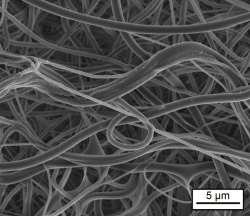
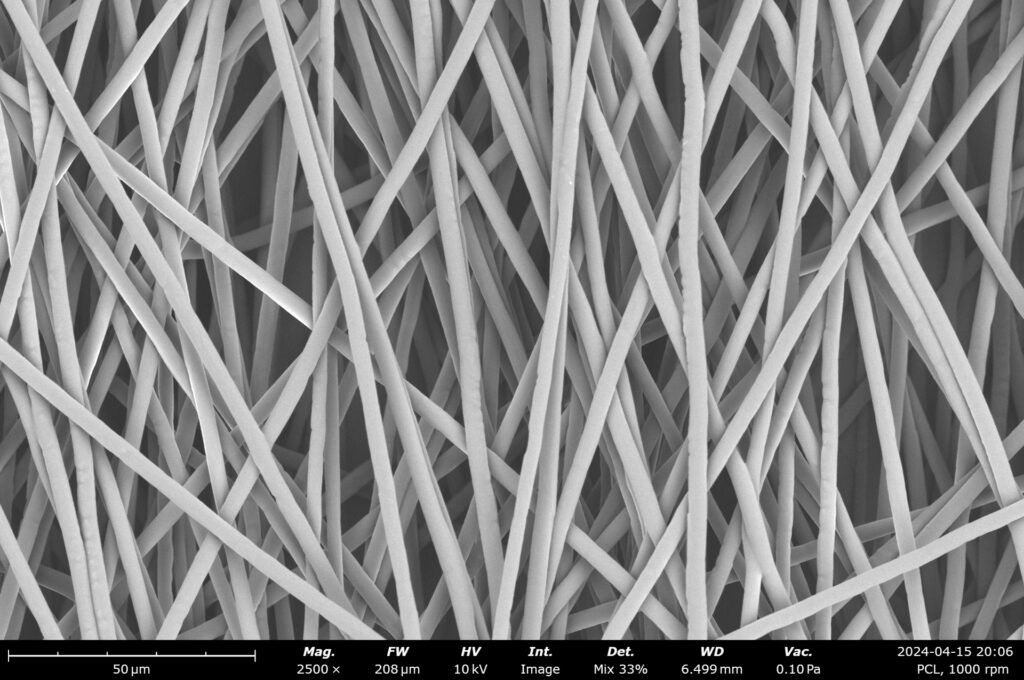
Microstructure of a tropoelastin nonwoven. Image: Fraunhofer IMWS

Electrospinning of Tropoelastin-Based Nanofibers

Electrospinning is widely used to fabricate electrospun biomaterials with nanoscale fibrous structures that mimic the architecture of the extracellular matrix. The technique involves the application of a high electric field to a polymer or protein solution, generating ultrafine fibers that are deposited as nonwoven membranes.

The electrospinning of tropoelastin has been studied as a method for producing porous protein scaffolds. Research has shown that the morphology of electrospun tropoelastin fibers can be modulated by varying spinning parameters such as delivery flow rate and starting protein concentration, and that cross-linked electrospun scaffolds retain the elasticity and cell-interactive properties inherent in the tropoelastin precursor (Wise, Mithieux & Weiss, 2009).

Electrospun scaffolds typically exhibit several characteristics that are advantageous for biomedical applications:

  • High surface-to-volume ratio
  • Porous fibrous architecture
  • Structural similarity to extracellular matrix networks
  • Tunable fiber diameter and scaffold morphology

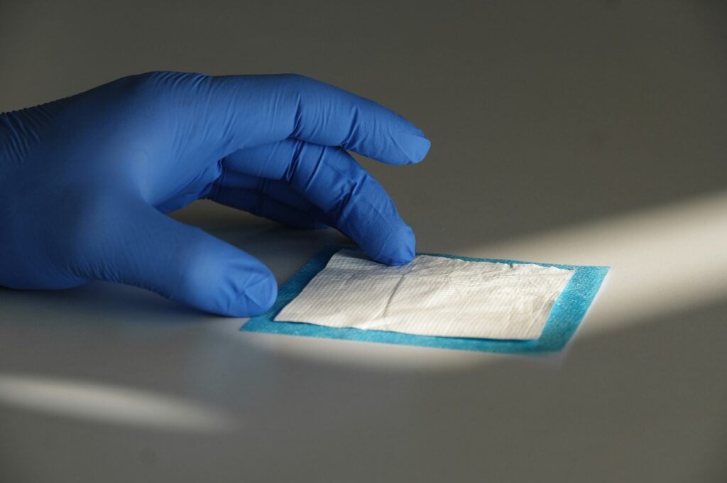
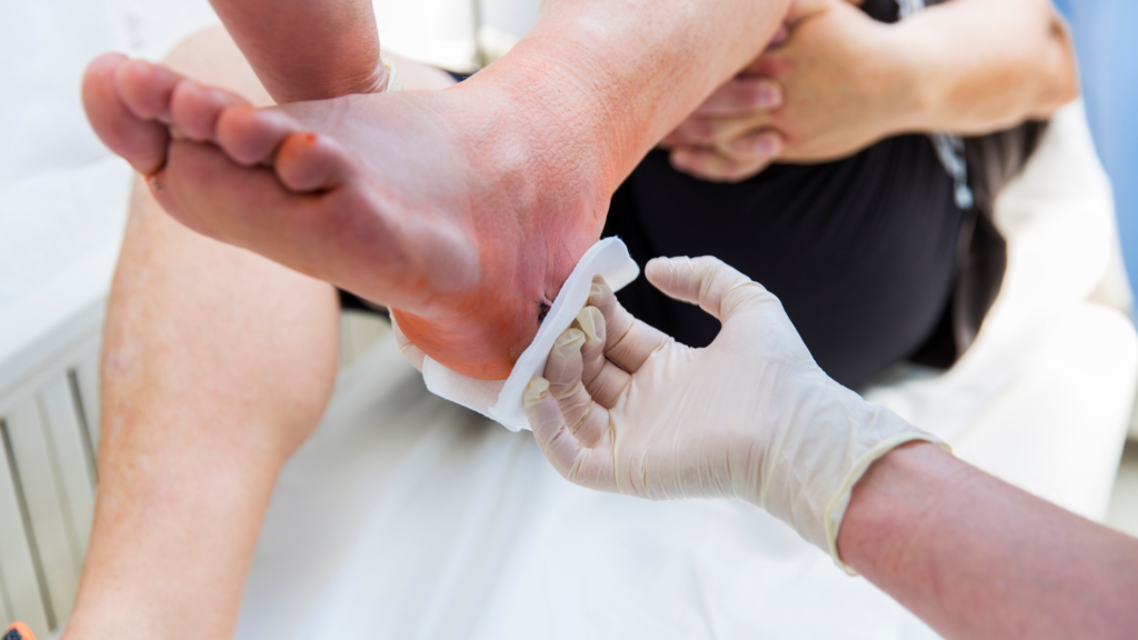
In the Fraunhofer-associated research project, tropoelastin-based materials are being developed as customized wound dressing materials, specifically targeting the treatment of chronic and complex wounds. These conditions represent a major challenge in healthcare, particularly in aging populations. Chronic wounds such as venous ulcers, leg ulcers, and foot ulcers often require long-term treatment and can significantly affect patients’ quality of life (Fraunhofer IMWS, 2024). These conditions are also associated with considerable healthcare costs. Preclinical tests described in the Fraunhofer IMWS project documentation indicate that the tropoelastin-based material developed within the collaboration is a promising candidate for use as a wound dressing in such contexts. The material combines biocompatibility, biodegradability, durability, and favorable mechanical behavior.

Microscopy Techniques for Electrospun Biomaterial Characterization

SEM and Microscopy Techniques for Nanofiber Characterization Analyzing the structural properties of electrospun biomaterials requires detailed nanofiber characterization techniques capable of resolving features at the micro- and nanoscale. Microscopy methods are essential tools in biomaterials research for this purpose.

Among the techniques commonly used in the field to analyze electrospun scaffolds are:

  • Scanning Electron Microscopy (SEM): provides high-resolution images of fiber networks, enabling evaluation of fiber uniformity, diameter distribution, and structural integrity.

Transmission Electron Microscopy (TEM): allows investigation of internal fiber structure and nanoscale features.

  • Atomic Force Microscopy (AFM): used to assess surface topography and nanomechanical properties of individual fibers.
  • Optical microscopy: applied for preliminary morphological assessment and scaffold-level organization.

Scanning Electron Microscopy is instrumental analysis for evaluating electrospun protein-based scaffolds, as it enables high-resolution visualization of nanofiber topographies. Through SEM-based morphometric analysis, critical parameters—including fiber diameter distribution, uniformity, and overall architecture can be quantified, providing essential insights into the materials performance within biological environments.

Fraunhofer IMWS Research on Elastin-Like Materials

The Fraunhofer Institute for Microstructure of Materials and Systems IMWS specializes in the structural characterization of advanced materials. Within the tropoelastin research project, the institute applies microscopy techniques to analyze the morphology and structural properties of the developed biomaterials (Fraunhofer IMWS, 2024).

By combining electrospinning with detailed microstructural characterization, the research team can evaluate how the processing of tropoelastin-based materials influences their final structural and mechanical properties. These analyses provide insights into key aspects such as:

  • Fiber morphology and structural organization
  • Scaffold architecture and fiber distribution
  • Surface morphology of the nanofibers
  • Potential relationships between microstructure and material performance
Electrospun nonwoven of biotechnologically produced tropoelastin

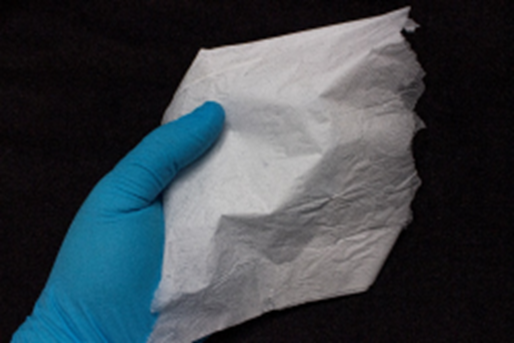
Electrospun nonwoven of biotechnologically produced tropoelastin. Image: Fraunhofer IMWS.

Material Properties of the Tropoelastin-Based Wound Dressing

The collaborative project aims to create customized wound dressings based on tropoelastin biomaterials. According to the Fraunhofer IMWS project description, the developed material demonstrates a combination of properties that are particularly relevant for wound care applications (Fraunhofer IMWS, 2024):

  • Biocompatibility
  • Biodegradability
  • Mechanical properties comparable to human skin in preclinical assessments
  • Durability suitable for biomedical use

These characteristics are essential for wound dressing materials intended to support tissue regeneration while maintaining mechanical compatibility with the surrounding tissue. An important aspect highlighted in the project documentation is the similarity between the mechanical properties of the tropoelastin-based material and those of human skin—a property that can be attributed to the role of elastin in conferring skin elasticity and resilience (Wise, Mithieux & Weiss, 2009). Microscopy characterization is central to evaluating these structural properties. By analyzing nanofiber morphology and scaffold architecture, researchers can assess whether the material structure supports its intended biomedical function. The detailed characterization results from the Fraunhofer IMWS project are expected to be published in peer-reviewed literature as the project progresses.

Implications for Regenerative Medicine and Biomedical Device Development

The treatment of chronic and complex wounds represents a significant medical challenge, particularly in aging populations. Conditions such as venous ulcers and foot ulcers often require long-term care and can lead to serious complications if not treated effectively (Fraunhofer IMWS, 2024).

Innovative biomaterials are therefore being actively investigated to improve wound healing outcomes. Materials that combine biocompatibility, mechanical compatibility with surrounding tissues, and structural similarity to natural ECM are particularly promising. Tropoelastin-based biomaterials represent one such approach: because tropoelastin is the monomer of elastin, materials derived from it can reproduce structural and mechanical characteristics relevant to skin and other elastic tissues (Almine et al., 2010).

The use of human-derived recombinant protein materials also addresses concerns associated with animal-derived biomaterials, including potential infection risks and immune responses. This is a recognized advantage in the development of next-generation protein biomaterials for clinical use (Wise, Mithieux & Weiss, 2009).

The integration of electrospinning technologies with advanced biomaterial characterization enables researchers to systematically investigate these materials. Through high-resolution microscopy and structural analysis, researchers can evaluate whether electrospun tropoelastin scaffolds exhibit the morphological and mechanical properties required for biomedical applications.

Such interdisciplinary approaches—combining biomaterials science, nanofiber fabrication, and microscopy analysis—are central to the development of next-generation biomaterials for regenerative medicine and wound care.

Conclusion

The development of customized wound dressings based on human tropoelastin represents a significant and scientifically grounded direction in biomaterials research. By leveraging the properties of the soluble elastin precursor, researchers aim to create biomaterials that replicate the elasticity and resilience of natural skin tissue.

The collaborative project involving Skinomics GmbH, Martin Luther University Halle-Wittenberg, and Fraunhofer IMWS highlights the importance of combining biomaterial design with advanced structural characterization. Microscopy analysis

plays a key role in understanding the fiber morphology and scaffold architecture of electrospun tropoelastin materials, ensuring they meet the requirements for biomedical applications.

Continued advancements in the characterization and fabrication of tropoelastin-based materials may transform the treatment for non-healing wounds, offering a biomimetic foundation for innovative tissue engineering applications.

Looking to Develop and Validate Innovative Biomaterials Like Tropoelastin?

Fluidnatek supports advanced electrospinning projects for the production of electrospun biomaterials and nanofiber scaffolds tailored for biomedical research and regenerative medicine applications. Whether your project involves protein-based materials, ECM-mimicking scaffolds, or wound care devices, our platforms are designed to support rigorous research in collaboration with institutions such as Fraunhofer IMWS.

Contact Fluidnatek to discuss how our electrospinning solutions can support your biomaterial development pipeline.

References

Almine, J. F., Bax, D. V., Mithieux, S. M., Nivison-Smith, L., Rnjak, J., Waterhouse, A., Wise, S. G., & Weiss, A. S. (2010). Elastin-based materials. Chemical Society Reviews, 39(9), 3371–3379. https://doi.org/10.1039/b919452p

Fraunhofer Institute for Microstructure of Materials and Systems IMWS. (2024). Innovative wound care – customized wound dressings made from tropoelastin [Project communication]. Fraunhofer IMWS. https://www.imws.fraunhofer.de

Mithieux, S. M., Wise, S. G., & Weiss, A. S. (2012). Tropoelastin – a multifaceted naturally smart material. Advanced Drug Delivery Reviews, 65(4), 421–428. https://doi.org/10.1016/j.addr.2012.06.009

Wise, S. G., & Weiss, A. S. (2009). Tropoelastin. International Journal of Biochemistry & Cell Biology, 41(3), 494–497. https://doi.org/10.1016/j.biocel.2008.03.017

Wise, S. G., Mithieux, S. M., & Weiss, A. S. (2009). Engineered tropoelastin and elastin-based biomaterials. Advances in Protein Chemistry and Structural Biology, 78, 1–24. https://doi.org/10.1016/S1876-1623(08)78001-5

Blit, P. H., Battiston, K. G., Yang, M., Paul Santerre, J., & Woodhouse, K. A. (2012). Electrospun elastin-like polypeptide enriched polyurethanes and their interactions with vascular smooth muscle cells. Acta Biomaterialia, 8(7), 2493–2503. https://doi.org/10.1016/j.actbio.2012.03.032

Electrospun Fibers as Implant Interface Layer: Modulating Implant–Tissue Interactions

Implant–Tissue

Introduction: Enhancing Implant–Tissue Interactions

The long-term performance of biomedical implants is fundamentally determined by the biological response at the implant–tissue interface. Regardless of the bulk material used—metallic, polymeric, or composite—the surface in contact with host tissue governs protein adsorption, immune activation, cellular adhesion, and ultimately tissue remodeling. Suboptimal interface properties can result in persistent inflammation, fibrous encapsulation, or postoperative adhesions, compromising both functional outcomes and patient recovery.

In recent years, electrospun nanofiber membranes have emerged as promising candidates for engineering implant interface layers. Their structural resemblance to the extracellular matrix (ECM), combined with tunable surface chemistry and degradability, enables controlled modulation of cell–material interactions.

A recent study by Ren et al. (2023) investigated electrospun polycaprolactone (PCL)/polyethylene glycol (PEG) membranes as implant interface layers. By varying PEG content, the authors tailored membrane hydrophilicity and evaluated its influence on macrophage response in vitro and adhesion formation in vivo using a rat Achilles tendon injury model. Surface hydrophilicity emerged as a key factor in attenuating inflammatory signaling and optimizing tissue-implant integration

This article examines electrospun fibers as implant interface layers, focusing on their biological rationale, material strategies, translational relevance, and fabrication considerations within the biomedical context.

What Are Implant Interface Layers and Why Do They Matter?

An implant-tissue interface serves as a functionalized interlayer or biomimetic scaffold engineered to modulate the bidirectional biological signaling between a prosthetic device and surrounding tissue. Evolving beyond inert coatings, these architectures now function as bioactive modulators that influence the acute immune response and subsequent long-term homeostatic integration

Key biological processes occurring at the implant interface include:

  • Adsorption of serum proteins
  • Recruitment and activation of immune cells
  • Macrophage polarization dynamics
  • Fibroblast migration and extracellular matrix deposition
  • Fibrotic encapsulation or adhesion formation

Macrophages play a central role in determining the fate of implanted materials. Their polarization toward a pro-inflammatory (M1-like) or pro-regenerative (M2-like) phenotype significantly influences healing outcomes. Excessive or prolonged M1 activation is associated with chronic inflammation and fibrosis, whereas M2 polarization supports tissue repair and remodeling.

In the study by Ren et al., bone marrow-derived macrophages (BMDMs) cultured on electrospun PCL/PEG membranes exhibited hydrophilicity-dependent responses. Increasing PEG content enhanced membrane hydrophilicity and was associated with down-regulation of inflammatory gene expression and increased expression of markers linked to M2-like polarization. These results demonstrate that surface wettability can meaningfully influence immune cell behavior.

In vivo evaluation using a rat model further demonstrated that pure PCL membranes were associated with substantial adhesion formation, whereas PCL/PEG membranes showed reduced adhesion and facilitated easier separation of tendon from surrounding tissue. The membrane containing the highest PEG ratio exhibited the lowest inflammatory response and fewest adhesions among the tested groups.

Electrospun nanofibers are thus repositioned as bioactive transducers capable of governing tissue-to-implant integration, moving beyond the concept of static anatomical barriers

Electrospun Nanofibers for Implant–Tissue Integration

Electrospinning produces continuous fibers with diameters typically in the nano- to submicron range, forming porous, interconnected membranes. Several characteristics make electrospun nanofibers particularly attractive as implant interface layers.

Advantages of Fibrous Biointerfaces

  1. ECM-Mimetic Architecture

The fibrous morphology of electrospun membranes resembles native extracellular matrix, providing topographical cues that influence cell adhesion and morphology. This structural similarity can facilitate more physiological cell–material interactions compared to smooth or minimally textured surfaces.

  1. High Surface Area and Porosity

Electrospun mats present large surface areas for protein adsorption and cell contact, while their interconnected porosity supports nutrient diffusion and cellular infiltration where desired.

  1. Tunable Surface Chemistry

By blending polymers with different physicochemical properties, such as hydrophobic PCL and hydrophilic PEG, membrane wettability and degradation behavior can be adjusted. In the Ren et al. study, increasing PEG content directly modulated hydrophilicity and altered macrophage responses.

  1. Controlled Degradation

The study noted that membranes with higher PEG content exhibited a sparser multilayer structure in vivo, which may be related to faster degradation and potentially facilitated tissue separation at the membrane layer. This observation suggests that degradation kinetics can influence adhesion formation and interface remodeling.

Materials Used and Functionalization Strategies

H3 PCL/PEG Blended Systems

Polycaprolactone (PCL) is a widely used biodegradable polyester known for its mechanical flexibility and slow hydrolytic degradation.

Nevertheless, its inherent hydrophobicity frequently leads to non-specific protein adsorption, which may trigger adverse pro-inflammatory responses.

Polyethylene glycol (PEG), in contrast, is hydrophilic and widely used to enhance surface wettability and reduce non-specific protein adsorption. By blending PEG with PCL prior to electrospinning, Ren et al. created membranes with tunable hydrophilicity while maintaining structural integrity.

The study demonstrates that increasing PEG content:

  • Enhances hydrophilicity
  • Reduces inflammatory gene expression in macrophages
  • Promotes M2-like polarization
  • Reduces adhesion formation in vivo

Importantly, the investigation did not rely on additional bioactive molecules or drug incorporation; the modulation effect was achieved solely through adjustment of polymer composition and resulting surface properties.


Role of Fiber Alignment

The membranes in the study were described as aligned nanofibers. Fiber alignment can influence cell orientation and migration, particularly in musculoskeletal applications where anisotropic tissue architecture is critical. While the study focuses primarily on hydrophilicity effects, alignment may contribute to guiding tissue organization at the interface.

Considerations for Surface Modification

Beyond polymer blending, electrospinning platforms allow additional strategies such as incorporation of bioactive agents or post-fabrication surface treatments. However, the Ren et al. work specifically highlights that even without complex biochemical functionalization, physicochemical modulation alone can significantly alter immune response and adhesion outcomes.

Applications and Clinical Relevance

Tendon Repair and Adhesion Prevention

Postoperative adhesions remain a major complication in tendon surgery, limiting mobility and functional recovery. In the rat Achilles tendon injury model used by Ren et al., pure PCL membranes were associated with substantial tissue adhesion. In contrast, PCL/PEG membranes reduced adhesion formation, and the highest PEG ratio yielded the most favorable outcome in terms of reduced inflammation and improved tissue separation.

These findings suggest that electrospun implant interface layers may serve as physical and biological barriers that minimize pathological fibrotic bridging while supporting controlled healing.


Broader Implications for Implant–Tissue Integration

Although the study specifically evaluates a tendon model, the underlying principle—modulation of macrophage phenotype through surface hydrophilicity—has broader implications for other soft tissue implant applications. However, extrapolation to orthopaedic hard-tissue implants or cardiovascular devices requires dedicated experimental validation.

The work supports a paradigm in which implant surface engineering prioritizes immune modulation as a primary design objective.

PCL aligned fibers made at 1000 rpm

PCL aligned fibers made at 1000 rpm. Image credit: Nanoscience Instruments.

Fluidnatek’s Capabilities in Implant Interface Nanofiber Development

Translating preclinical findings into practical biomedical applications requires reproducible fabrication platforms capable of controlling fiber morphology, alignment, and polymer composition.

Fluidnatek provides electrospinning systems designed to support:

  • Precise control of polymer blending (e.g., PCL/PEG ratios)
  • Fabrication of aligned nanofiber membranes
  • Reproducible control over fiber diameter and morphology
  • Development of degradable fibrous interface layers

Such platforms enable research teams to replicate and extend experimental configurations similar to those described by Ren et al., facilitating systematic studies on implant–tissue integration and immune modulation.

More information on electrospinning platforms for biomedical research is available at: https://fluidnatek.com/electrospinning-machines/

Conclusion: Toward Immunomodulatory Implant Interfaces

Electrospun fibers as implant interface layers represent a strategic evolution in biomedical surface engineering. Rather than functioning solely as passive structural coatings, these nanofiber membranes can actively influence early immune responses and subsequent tissue remodeling.

The study by Ren et al. demonstrates that tuning hydrophilicity through PCL/PEG blending modulates macrophage gene expression and reduces adhesion formation in a rat tendon injury model. Increased PEG content correlated with reduced inflammatory signaling, enhanced M2-like polarization, and fewer postoperative adhesions. Additionally, higher PEG ratios were associated with structural changes consistent with faster degradation, which may facilitate tissue separation at the interface.

These findings reinforce the concept that surface chemistry and nanoscale architecture are central determinants of implant performance. Continued investigation into electrospun nanofiber interface layers may advance the development of next-generation biomedical implants designed not only for mechanical function, but also for precise biological integration.

References

Ren, Y., et al. (2023). Electrospun fibers as implant interface layer. ElectrospinTech. Retrieved from http://electrospintech.com/implantinterface.html

Zhang, X., Liu, L., Wang, Y., & Chen, H. (2021). Electrospun nanofiber scaffolds in regenerative medicine. Acta Biomaterialia, 134, 123–140. https://doi.org/10.1016/j.actbio.2021.04.010

Li, Q., Yang, J., Zhao, Y., & Wang, L. (2020). Electrospun nanofibers as implant coatings for tissue regeneration. Journal of Biomedical Materials Research Part A, 108(9), 1834–1845.


Case Study — Evonik & VECOLLAN®: Recombinant Collagen Nanofiber Manufacturing Through Electrospinning with Fluidnatek® LE-50

VECOLLAN Fluidnatek

Animal-Free Alternatives in Biomedical Materials

The biomedical sector is undergoing a decisive transition toward fully animal-free materials for regenerative medicine, advanced wound care, and premium cosmetic technologies. This shift is driven not only by ethical considerations but also by growing regulatory requirements for full traceability, pathogen safety, and reproducible manufacturing processes.

In this context, Evonik has developed VECOLLAN®—a recombinant collagen-like peptide designed for biomedical applications. VECOLLAN® is produced through a scalable, reproducible fermentation-based process and offers exceptional purity, safety, and consistency.

In a recent study, Evonik utilized VECOLLAN® to create electrospun meshes using the Fluidnatek® LE-50 equipment—a versatile electrospinning platform for advanced research and pilot-scale process optimization. The LE-50 enabled a coaxial electrospinning setup, placing VECOLLAN® in the fiber core while distributing a controlled crosslinking agent in the outer shell. This configuration delivered three key benefits:

  • Enhanced mechanical stability of the scaffold
  • Reduced swelling in biological environments
  • Tunable dissolution behavior

These properties are critical for implantable devices, controlled drug-release platforms, and next-generation wound care solutions.

This case study demonstrates how Fluidnatek® systems empower the development of next-generation biomaterials—consistent, safe, sustainable. The LE-50’s flexibility, environmental control, and compatibility with post-processing integrations make it an essential tool for organizations seeking to accelerate innovation while minimizing process risk and time to market.

👉 Official Evonik publication: Recombinant collagen platforms 

  1. Krauss C, Montero Mirabet M, Zhang JF, Mader K. Electrospinning of animal-free derived collagen-like protein: Development and characterization of VECOLLAN(R)- nanofibers for biomedical applications. Int J Pharm X. 2025;10:100398.

Fluidnatek Strengthens Its Commitment to Biomedical Innovation at COMPAMED 2025

Fluidnatek COMPAMED 2025

Fluidnatek successfully participated in MEDICA-COMPAMED 2025, the leading international event for the healthcare industry, which brought together over 5,300 exhibitors from 70 nations and attracted 78,000 professional visitors from November 17 to 20 in Düsseldorf. This participation provided a valuable opportunity to connect with the international scientific community and gain deeper insights into the trends shaping the future of biomedical applications.

A Strategic Encounter with the Global Healthcare Ecosystem

From Stand 8bK34 in Hall 8B at COMPAMED, our team conducted live demonstrations of the LE-50 Gen2 system throughout all four days of the fair, allowing visitors to experience firsthand the capabilities of electrospinning technology and establish meaningful connections with top-level professionals in the sector. The fair, which adopted the theme “Meet Health. Future. People.” this year, consolidated its position as an essential meeting point for healthcare industry decision-makers. According to the organizers’ data, three-quarters of professional visitors belong to senior management at their companies or organizations, and 75% traveled from 160 different countries, confirming the truly global reach of the event.

The intensive days in Düsseldorf proved particularly enriching for Fluidnatek. The dynamic exchanges with visitors from different regions around the world provided valuable perspectives on current challenges in the biomedical sector and emerging needs in areas such as tissue engineering, regenerative medicine, and advanced drug delivery systems.

Key Learnings for Future Development

Participation in MEDICA-COMPAMED 2025 enabled Fluidnatek to identify important trends that will guide our technological development in the coming years:

Tissue Regeneration and Personalized Medicine: Conversations with researchers and clinical professionals revealed a growing demand for more versatile solutions for creating 2D and 3D scaffolds tailored to specific applications, from bone and cartilage regeneration to vascular engineering.

Advanced Wound Healing: The interest shown in next-generation wound dressings with superior healing properties underscores the need to continue innovating in functional materials that integrate antimicrobial capabilities, growth factors, and controlled release of active ingredients.

Smart Medical Devices: The integration of specialized coatings in medical devices with complex geometries emerges as a high-potential area, particularly in implants and devices with prolonged tissue contact.

Controlled Release Platforms: The development of drug delivery systems based on functionalized nanofibers remains a field of great interest, particularly in oncology, chronic disease treatment, and localized therapies.

Strategic Collaborations and Industry Synergies

One of the most valuable aspects of participating in COMPAMED has been the opportunity to establish dialogues with leading companies in the sector.
This environment has allowed Fluidnatek to position itself as a technology partner specializing in electrospinning and electrospraying processes, with capabilities ranging from biomedical research to applications in pharmacy, cosmetics, filtration, energy, and new materials.

Looking Toward the Future of Biomedicine

The experience at MEDICA-COMPAMED 2025 reinforces Fluidnatek’s vision of the transformative role that nanofiber technologies can play in the medicine of the future. The conversations held during the fair provided valuable insights into the directions in which the biomedical sector is evolving:

  • The growing demand for solutions for organoids and complex tissue models that enable advances in personalized medicine and more predictive preclinical trials.
  • Interest in sterile applications and systems that ensure maximum safety for implants and devices in direct contact with the organism.
  • The need for scalability and reproducibility in the manufacturing of advanced biomedical materials.
  • The integration of multiple functionalities into a single technological platform, combining mechanical, biological, and pharmacological properties.


COMPAMED_booth

Becky Thunio and Enrique Navarro at the Fluidnatek booth during COMPAMED 2025.

Ongoing Commitment to Innovation

The next edition of MEDICA and COMPAMED will take place from November 16 to 19, 2026, in Düsseldorf. The organizers have announced they will continue developing both events toward greater integration, leveraging synergies and expanding their international relevance, with the goal of facilitating even more intensive interdisciplinary dialogue among industry, science, politics, and clinical practice.

For Fluidnatek, participation in MEDICA-COMPAMED is not simply an exhibition opportunity, but an ongoing commitment to learning, collaborative innovation, and developing solutions that respond to the real needs of the biomedical sector. The knowledge acquired at this edition will guide our R&D efforts and allow us to remain a reference in electrospinning technologies for the advancement of biomedical applications.

We thank all the professionals who visited our stand and shared their experiences and visions about the future of biomedicine. These exchanges are fundamental to continuing the development of technologies that truly make a difference in people’s health and well-being.

Fluidnatek at DGBM 2025: Shaping the Future of Biomedical Materials

The German Society for Biomaterials 2025 (DGBM) conference in Dresden has wrapped up, leaving us inspired and grateful for the vibrant exchange of knowledge with leading experts in biomaterials and regenerative medicine.

A heartfelt thank you to the DGBM organization for hosting such an impactful event and to every delegate who contributed to deep discussions around the future of electrospun nanofibers and their role in innovative therapies and advanced drug delivery.

Fluidnatek is proud to strengthen its positioning in the biomedical community and to continue revolutionizing nanofiber solutions with cutting-edge electrospinning technology. Special thanks to our colleagues Becky Tunio (KAM) and Enrique Navarro (Sales & Marketing Manager) for representing our commitment and expertise on-site.

Let’s keep pushing the boundaries of innovation together!

More about the event: https://www.dgbm-kongress.de/

Becky Tunio and Enrique Navarro Alonso, at DGBM 2025.

Fluidnatek Unveils Revolutionary LE-50 Gen2: Next-Gen Biomedical Innovation Takes Center Stage at Medical Technology Ireland 2025

2025 MTI

Fluidnatek made a significant impact at Medical Technology Ireland 2025, held September 24–25 at the Galway Racecourse, where we proudly unveiled our groundbreaking LE-50 Gen2 electrospinning and electrospraying platform. This cutting-edge system represents the future of nanofiber and nanoparticle research in biomedical applications.

Live Innovation in Action

Our exhibition stand became a hub of scientific discovery as attendees witnessed live demonstrations of the LE-50 Gen2‘s remarkable capabilities. This state-of-the-art benchtop system revolutionizes laboratory research by seamlessly integrating both needle-based and needleless technologies within a single, versatile unit.

Key breakthrough features include:

  • Dual-solution processing capabilities
  • Independent high-voltage control systems
  • Automated emitter motion ensuring exceptional homogeneity
  • Unmatched precision for multi-material scaffold development

These advanced functionalities position the LE-50 Gen2 as the ideal solution for pioneering applications in tissue engineering, accelerated wound healing, precision drug delivery systems, and next-generation medical device coatings.

Expert Representation

Fluidnatek’s presence was expertly represented by our specialized team:

  • Enrique Navarro, Sales & Marketing Manager
  • Milan Proks, Key Account Manager

Transforming Medical Science

Electrospinning technology is revolutionizing biomedical research by enabling the creation of nanofiber-based scaffolds that precisely replicate the natural extracellular matrix. This biomimetic approach significantly enhances cell growth and accelerates tissue regeneration processes. Additionally, our electrospun materials deliver controlled, targeted release of therapeutic compounds, opening new frontiers in personalized medicine.

The LE-50 Gen2’s exceptional precision combined with its scalability makes it an indispensable tool for researchers and companies driving the next wave of medical technology breakthroughs.

Looking Forward

We extend our sincere gratitude to all the innovators, researchers, and industry leaders who visited our stand and engaged in meaningful discussions about how Fluidnatek’s advanced solutions can accelerate biomedical innovation. These valuable conversations fuel our commitment to pushing the boundaries of what’s possible in medical technology.

For more information about the LE-50 Gen2 and how it can transform your biomedical research, contact our team today.

2025 MTI

Live demonstrations of the LE50 Gen2.

Engaging with the Biomedical Community at FBPS 2025 in Porto

FBPS Porto

Showcasing innovation in electrospinning and biomedical polymers

Fluidnatek successfully participated in the FBPS 2025 – Biomedical Polymers & Electrospinning Symposium, recently held in Porto. This international symposium provided a unique opportunity to present our latest innovations in electrospinning technology, nanofibers for biomedical applications, and advanced polymers, while strengthening collaboration with the global scientific community.

Event highlights

Innovative solutions on display

We showcased our latest developments in nanofiber electrospinning, nanotechnology, and biomedical applications, attracting strong interest from researchers and industry professionals.

Knowledge exchange

Our team engaged with international experts, generating enriching discussions and potential collaborations for future projects in biomaterials and nanofibers.

Excellent reception at our booth

Many visitors approached our booth to learn more about our technology, explore applications, and discuss opportunities for scientific and industrial collaboration.

Looking ahead

We would like to thank the symposium organizers for such an inspiring edition, as well as all visitors who shared their ideas and enthusiasm with us.

Events like FBPS 2025 confirm that we are on the right path: continuing to innovate in electrospinning, strengthen ties with the scientific community, and develop solutions with a real impact in biomedical applications.

Discover more about our electrospinning technologies and how we apply nanofibers and advanced polymers in biomedicine.

FBPS25_Becky

Becky Tunio, at FBPS 2025 in Porto.

Electrospun Bioresorbable Tubular Scaffolds for Advanced Medical Devices

electrospun bioresorbable tubular scaffolds

Introduction: The Need for Biofunctional Medical Devices

Electrospinning has emerged as a transformative technology in biomedical engineering, enabling the fabrication of nanofibrous materials that closely mimic the hierarchical structure and functionality of the extracellular matrix (ECM) found in native tissues. This biomimetic capability is particularly valuable for developing next-generation medical devices including vascular grafts, stent coatings, bioresorbable stents, nerve conduits, and electrospun bioresorbable tubular scaffolds. These applications demand precise control over material architecture, mechanical properties, biocompatibility, and degradation kinetics to achieve optimal functional performance.

The growing demand for minimally invasive, patient-specific interventions has accelerated interest in electrospun tubular constructs that can be fully resorbed by the body after fulfilling their therapeutic function. This application note explores the current state of electrospinning technology for producing electrospun bioresorbable tubular scaffolds, highlights key applications in medical device development, and discusses emerging trends in this rapidly evolving field.


Electrospinning Technology for Bioresorbable Tubular Scaffold Production

Process Fundamentals

Electrospinning for tubular scaffold fabrication involves applying a high voltage (10-30 kV) to a polymer solution or melt, creating an electrostatic force that overcomes surface tension to form a jet. This jet undergoes whipping and stretching as the solvent evaporates, resulting in nanofibers that collect on a opposite voltage rotating mandrel to form tubular structures. The process allows precise control over:

  • Fiber diameter (typically 100-500 nm)
  • Fiber orientation (random or aligned)
  • Porosity (60-90%)
  • Wall thickness (50 μm to several mm)
  • Mechanical properties (tensile strength, compliance, and elasticity)
  • Surface chemistry and topography

Equipment Configurations

Several commercial systems have been developed specifically for tubular scaffold production, including the Fluidnatek LE-100 Bio Tubing platform. These advanced electrospinning systems typically feature:

  • Multiple collector options: Rotating mandrels with variable diameters (0.5-10 mm) and rotation speeds (50-2000 rpm) for seamless tubular scaffold fabrication
  • In-line monitoring: Real-time thickness measurement and fiber morphology analysis for stringent quality control
  • Environmental control: Precision regulation of temperature (18-45°C) and humidity (10-80% RH) to ensure reproducibility
  • Clean processing environments: ISO 5/Class 100 compatible chambers for aseptic, contamination-free processing
  • Automation capabilities: Programmable deposition patterns and process parameters for complex architectures
  • Data management: Industry 4.0 integration for process traceability and validation

Materials for Electrospun Bioresorbable Scaffolds

The selection of appropriate polymers is critical for successful bioresorbable scaffold development. Commonly used materials include:

Polymer

Degradation Time

Key Properties

Common Applications

Poly(lactic acid) (PLA)

12-24 months

High strength, moderate hydrophobicity

Vascular grafts, bone scaffolds

Poly(glycolic acid) (PGA)

2-4 months

Rapid degradation, good cell adhesion

Nerve guides, temporary stents

Poly(lactic-co-glycolic acid) (PLGA)

1-12 months (tunable)

Controllable degradation rate

Drug delivery, soft tissue engineering

Polycaprolactone (PCL)

24-36 months

Excellent elasticity, slow degradation

Long-term vascular applications

Polyurethanes (PU)

Variable

Superior mechanical properties

Heart valves, vascular devices

Natural polymers (collagen, silk, chitosan)

Variable

Enhanced bioactivity

Tissue engineering, wound healing

Multi-material approaches using polymer blends or core-shell configurations enable tailored degradation profiles and mechanical properties specific to each application.

Electrospun Scaffolds for Medical Devices and Tissue Engineering

Electrospun bioresorbable tubular scaffolds are advancing several areas in medical device development:

Electrospun Vascular Grafts

Electrospun vascular grafts represent a promising alternative to autologous vessels for bypass procedures and vascular repair. Their advantages include:

  • Tunable compliance: Matching mechanical properties with native vessels reduces hemodynamic disturbances and intimal hyperplasia
  • Controlled porosity: Optimized pore size (typically 10-30 μm) facilitates cell infiltration while maintaining barrier function
  • Drug delivery capabilities: Incorporation of anticoagulants, anti-inflammatories, or growth factors enhances performance
  • Degradation synchronized with tissue regeneration: Scaffold provides initial support and gradually transfers load to newly formed tissue

Clinical studies have demonstrated promising results for small-diameter (<6 mm) vascular grafts, with ongoing trials for peripheral and coronary applications.

Stent Coatings and Fully Bioresorbable Stents

Electrospun polymeric coatings for conventional metal stents (including nitinol-based stents) as well as fully bioresorbable stent platforms offer several advantages:

  • Controlled drug elution: Precise release kinetics for antiproliferative agents
  • Reduced foreign body response: Gradual dissolution minimizes chronic inflammation
  • Preservation of vessel geometry: After resorption, native vessel mechanics are restored
  • Facilitation of repeat interventions: Absence of permanent implants simplifies future procedures
  • Enhanced compatibility with nitinol stents: Electrospun coatings can mitigate nickel ion release while maintaining the mechanical advantages of nitinol.

Recent innovations include dual-layer electrospun stents with different drug release profiles and mechanical properties in each layer[8].

Nerve Conduits and Neural Tissue Engineering

Tubular electrospun conduits support nerve regeneration following injury by:

  • Directing axonal growth: Aligned nanofibers guide regenerating neurons
  • Preventing scar tissue infiltration: Semipermeable walls block fibroblast migration
  • Supporting Schwann cell migration: Optimized architecture promotes cellular colonization
  • Delivering neurotrophic factors: Sustained release of growth factors enhances nerve regeneration

Electrospun nerve guides have shown promising results in peripheral nerve defects up to 30 mm in preclinical models.

Hybrid Metal-Polymer Scaffolds

An important advancement in electrospun scaffold technology is the development of hybrid constructs combining metallic frameworks with electrospun polymer coatings. Nitinol (nickel-titanium alloy) is particularly valuable in these applications due to its unique properties:

  • Shape memory effect: Allows for minimally invasive deployment and self-expansion
  • Superelasticity: Provides mechanical support while maintaining flexibility
  • Biocompatibility: Well-established safety profile in vascular applications
  • Fatigue resistance: Withstands physiological cyclic loading

Electrospun coatings on nitinol structures can:

  • Deliver therapeutic agents locally
  • Modulate the tissue-material interface
  • Provide a template for tissue ingrowth
  • Create a barrier to control nitinol ion release

These hybrid constructs are particularly valuable for stents, occlusion devices, and embolic protection systems where the mechanical properties of nitinol complement the biological functionality of electrospun polymers[10].

Other Emerging Applications

Additional applications leveraging electrospun bioresorbable tubular scaffolds include:

  • Tracheal and bronchial replacement: Reinforced electrospun constructs with radial rigidity and longitudinal flexibility
  • Gastrointestinal stents: Degradable stents for temporary stricture management
  • Urethral reconstruction: Tailored scaffolds supporting regeneration of functional urethral tissue
  • Drug delivery conduits: Tubular implants for localized, sustained therapeutic delivery

Manufacturing Considerations

Quality Control Parameters

Consistent performance of electrospun tubular scaffolds depends on rigorous quality control focused on:

  • Structural uniformity: Even fiber distribution and orientation throughout the scaffold
  • Mechanical consistency: Batch-to-batch reproducibility of tensile strength, burst pressure, and compliance
  • Chemical purity: Residual solvent levels below regulatory thresholds (<50 ppm for common solvents)
  • Sterility assurance: Validated sterilization processes compatible with delicate nanostructures

Scale-Up Strategies

Transitioning from laboratory to commercial production requires addressing several challenges:

  • Throughput enhancement: Multinozzle or needleless systems to increase production volume
  • Process validation: Design of Experiments (DoE) approaches to establish robust process parameters
  • Inline monitoring: Real-time quality verification systems for continuous production
  • Regulatory compliance: Documentation systems meeting cGMP, ISO 13485, and FDA requirements
  • Sterilization compatibility: Process development for terminal sterilization methods preserving scaffold integrity

Regulatory Considerations

Electrospun bioresorbable scaffolds face specific regulatory challenges:

  • Novel material combinations: May require additional biocompatibility and degradation testing
  • Long-term degradation products: Assessment of tissue response to breakdown components
  • Process validation: Critical process parameters for electrospinning must be thoroughly documented
  • Mechanical testing standards: Often requires development of custom test methods specific to the intended application
  • Shelf-life determination: Stability of both mechanical properties and biological activity must be demonstrated

Regulatory pathways differ by region and specific application, with combination products (incorporating drugs or biologics) facing more complex requirements.

Clinical Case Studies

Case Study 1: Small-Diameter Vascular Grafts

A recent clinical trial evaluated PCL/PLA electrospun grafts (4 mm diameter) for hemodialysis access in 12 patients. Key findings included:

  • 83% primary patency at 6 months
  • No aneurysm formation or catastrophic mechanical failure
  • Progressive endothelialization observed via ultrasound
  • Degradation profile matching tissue ingrowth rates

Case Study 2: Drug-Eluting Bioresorbable Stent Coating

A PLGA electrospun coating on a metal stent platform demonstrated:

  • Reduced restenosis rates compared to bare metal stents (8% vs. 22%)
  • Complete resorption by 9 months post-implantation
  • Reduced dual antiplatelet therapy requirements
  • Improved vessel healing and reduced inflammation

Future Trends and Challenges

Several emerging approaches are poised to advance electrospun tubular scaffold technology:

  • Smart responsive scaffolds: Integration of stimuli-responsive materials that adapt to physiological changes
  • 4D printing approaches: Electrospun structures programmed to change shape or properties over time
  • Hybrid manufacturing: Combining electrospinning with other fabrication techniques (3D printing, textile processes)
  • Cell electrospinning: Direct incorporation of living cells during the fabrication process
  • Personalized medicine applications: Patient-specific scaffold designs based on medical imaging data

Challenges requiring further research include:

  • Mechanical property optimization: Matching complex native tissue mechanics more precisely
  • Control of degradation heterogeneity: Ensuring uniform resorption throughout the scaffold volume
  • Scale-up limitations: Addressing throughput constraints for high-volume applications
  • Standardization: Developing consensus testing methods specific to electrospun materials


Conclusion

Electrospun bioresorbable tubular scaffolds represent a significant advancement in medical device technology, offering unprecedented control over scaffold architecture, material properties, and biological response. As manufacturing capabilities continue to mature and clinical evidence accumulates, these materials are positioned to address critical unmet needs in vascular, neural, and other tubular tissue applications. Continued innovation in materials, processing techniques, and hybrid approaches will further expand the potential of this versatile technology platform.

Designed for Excellence in Tubular Scaffold Manufacturing
The Fluidnatek LE-100 BioTubing system is specially engineered to meet the stringent requirements of tubular scaffold production. Its advanced rotating mandrel system, precision-controlled environment, and high-resolution deposition capabilities enable the fabrication of seamless, uniform, and reproducible tubular structures. With full GMP-compliant architecture and options for cleanroom integration, the LE-100 BioTubing is the ideal platform for scaling up from research to clinical manufacturing of bioresorbable vascular grafts, nerve conduits, and other implantable devices.

Let’s Build the Future of Medical Devices
Are you developing resorbable scaffolds for advanced biomedical applications

**Fluidnatek’s electrospinning platforms** deliver the precision, reproducibility, and scalability needed to design **customised tubular nanostructures** for next-generation medical devices. 

👉 Contact our team (https://fluidnatek.com/contact) to discuss your biomedical project.

References

  1. Zhang Y, et al. Recent advances in electrospinning for biomedical applications. Biomater Sci. 2022;10(2):316-339. https://doi.org/10.1039/D1BM01518C
  2. Sensini A, et al. Hierarchical electrospun tendon-ligament bioinspired scaffolds. Biofabrication. 2023;15(1):015004. https://doi.org/10.1088/1758-5090/aca8c6
  3. Keirouz A, et al. Nanofiber-based wound dressings and their applications. Mater Sci Eng C. 2023;113:111018. https://doi.org/10.1016/j.msec.2020.111018
  4. Khorshidi S, et al. A review of key challenges of electrospun scaffolds for tissue-engineering applications. J Tissue Eng Regen Med. 2022;16(3):195-215. https://doi.org/10.1002/term.3267
  5. Gao S, et al. Core-shell nanofibers: Nano channel and capsule by coaxial electrospinning. Adv Mater Interfaces. 2023;10(7):2101770. https://doi.org/10.1002/admi.202101770
  6. Nagarajan S, et al. Design strategies for controlling degradation rate and mechanical properties in electrospun vascular scaffolds. ACS Appl Mater Interfaces. 2022;14(41):45829-45843. https://doi.org/10.1021/acsami.2c09274
  7. Fukunishi T, et al. Tissue-engineered small diameter arterial vascular grafts from cell-free nanofiber PCL/chitosan scaffolds in a sheep model. PLoS One. 2022;17(3):e0254315. https://doi.org/10.1371/journal.pone.0254315
  8. Qiu X, et al. Controlled dual-drug release from electrospun nanofibers as bioresorbable local drug delivery systems. J Control Release. 2023;353:607-618. https://doi.org/10.1016/j.jconrel.2022.12.039
  9. Wang S, et al. Aligned electrospun polycaprolactone/silk fibroin core-shell nanofibers for nerve tissue engineering. J Biomed Mater Res A. 2023;111(5):814-826. https://doi.org/10.1002/jbm.a.37487
  10. Torres-Giner S, et al. Industrial applications of electrospinning: Drug delivery, tissue engineering, and regulatory considerations. Int J Mol Sci. 2023;24(4):3676. https://doi.org/10.3390/ijms24043676
  11. Tsetsekou M, et al. Nitinol-polymer composites for medical applications: A review. J Mater Sci. 2023;58(10):4692-4721. https://doi.org/10.1007/s10853-022-08128-1
  12. Kuznetsov K, et al. Surface modification of nitinol stents with electrospun bioresorbable polymers: Approaches and clinical outcomes. J Biomater Appl. 2022;37(3):481-496. https://doi.org/10.1177/08853282221131975

Electrospun Wound Dressing: A Breakthrough in Advanced Wound Healing

wound-dressing-electrospinning

Electrospinning has emerged as a transformative technology for designing next-generation wound dressings. The unique ability of this technique to produce nanofiber-based scaffolds that mimic the extracellular matrix (ECM) has positioned it at the forefront of biomedical research. As chronic wounds, burns, and post-surgical injuries demand increasingly sophisticated care, electrospun wound dressings offer unmatched potential for accelerating healing, preventing infections, and delivering therapeutic agents in a controlled manner.

The Clinical Challenge in Wound Care

Chronic and acute wounds remain a significant clinical burden, particularly among aging populations and individuals with diabetes, vascular disease, or immunocompromised states. Conventional dressings often fail to provide optimal moisture retention, mechanical protection, or antimicrobial activity. Furthermore, they rarely support cellular activities required for tissue regeneration.

In contrast, nanofiber wound dressing systems can be engineered to address these limitations through structural mimicry of native tissue, functional loading with bioactive compounds, and controlled drug release. The growing body of research and innovation in biomedical electrospinning highlights the urgent need for advanced wound dressing materials.

human skin wound

View of a human skin wound.

Benefits of Electrospun Nanofibers for Wound Care

Electrospinning enables the production of continuous fibers with diameters ranging from tens of nanometers to a few micrometers, offering several biomedical advantages:

Mimicking the Extracellular Matrix (ECM)

The fibrous architecture of electrospun mats closely resembles the ECM, providing a favorable environment for cell adhesion, proliferation, and differentiation. This promotes effective re-epithelialization and granulation tissue formation.

Tunable Porosity and Moisture Control

By adjusting parameters such as voltage, flow rate, and polymer concentration, the porosity of the electrospun membrane can be finely tuned. This facilitates gas exchange while preventing bacterial infiltration, which is vital for wound healing.

Functionalization with Bioactive Agents

Nanofiber scaffolds can be functionalized with antimicrobial agents, growth factors, and anti-inflammatory drugs, enabling drug-loaded electrospun fibers that actively participate in the healing process rather than serving as passive barriers.

Mechanical Adaptability

Electrospun mats can be designed with elasticity and strength suitable for various anatomical sites, from joints to pressure points, enhancing patient comfort and compliance.


Polymeric Systems and Functionalization Strategies

The choice of polymers significantly influences the properties and functionality of electrospun wound dressings. Both synthetic and natural polymers are employed, often in blends to balance biocompatibility, degradability, and mechanical performance.

Synthetic Polymers for Structural Integrity

Polymers such as polycaprolactone (PCL), poly(lactic acid) (PLA), and polyurethane (PU) are frequently used due to their mechanical robustness and processability. These materials ensure the scaffold maintains structural integrity over time.

Biopolymers for Antimicrobial Effect and Bioactivity

Natural polymers, including collagen, gelatin, chitosan, and hyaluronic acid, offer inherent bioactivity. Biopolymer wound dressing systems leverage these materials to introduce antimicrobial and hemostatic properties.

For instance, chitosan is widely recognized for its antimicrobial properties and has been incorporated into nanofibrous matrices to enhance wound healing efficacy PubMed source.


Drug Delivery and Bioactive Capabilities

Electrospinning facilitates controlled drug release by embedding pharmaceuticals within or on the surface of the nanofibers. This delivery mode ensures a sustained release at the wound site, improving therapeutic outcomes and reducing systemic side effects.

Release Kinetics and Porosity Design

By modulating the polymer composition and fiber morphology, researchers can customize release profiles ranging from burst release to prolonged delivery over several days or weeks. Porosity design plays a critical role in mediating this process and can be optimized for different wound types and stages.

Multi-drug and Layered Systems

Advanced configurations such as core–shell nanofibers, multilayered mats, and coaxial spinning enable incorporation of multiple drugs with staggered release kinetics. This is especially valuable in treating infected wounds or those requiring both antimicrobial and regenerative agents.

Examples include loading electrospun mats with silver nanoparticles for antibacterial effects alongside vascular endothelial growth factor (VEGF) for tissue regeneration ScienceDirect source.

Vascular endothelial growth factor A (VEGF A) protein molecule

Vascular endothelial growth factor A (VEGF A) protein molecule. Cartoon representation combined with semi transparent surfaces.

Clinical Potential and Future Perspectives

The translation of electrospinning for biomedical applications from bench to bedside is accelerating. Several preclinical studies and early-stage clinical trials highlight the promising outcomes of wound healing scaffolds based on electrospun materials.

Regulatory Considerations

Despite the promise, regulatory hurdles persist. Sterilization techniques, reproducibility of fiber architecture, and scalability for mass production are key challenges. However, platforms like Fluidnatek® electrospinning systems are designed to meet Good Manufacturing Practice (GMP) requirements, easing the path to commercialization.

Personalized and Smart Dressings

Future directions point toward personalized wound care solutions, integrating biosensors for real-time monitoring, stimuli-responsive drug release, and AI-assisted design of scaffold parameters based on wound morphology.

Innovative research in wound healing biomaterials is increasingly leveraging machine learning and big data analytics to fine-tune material properties for individualized therapy.


Conclusion: From Research to Clinical Application

Electrospun wound dressings are reshaping the landscape of wound management. Their unique combination of biomimetic structure, bioactivity, and versatility makes them ideal candidates for a wide range of clinical applications—from diabetic ulcers to battlefield injuries.

As the field progresses, the synergy between material science, bioengineering, and medical practice will drive the development of even more effective solutions.

Are you exploring advanced wound care materials? Discover how Fluidnatek’s electrospinning platforms help design, test and scale biomedical nanofiber dressings tailored to your research or product needs. Explore our biomedical electrospinning solutions.


References

  1. Chouhan, D., & Mandal, B. B. Silk biomaterials in wound healing and skin regeneration therapeutics: From bench to bedside. Acta Biomaterialia, 2020, 103, 24–51. DOI: 10.1016/j.actbio.2019.11.050
  2. Boateng, J. S., Matthews, K. H., Stevens, H. N. E., & Eccleston, G. M. Wound healing dressings and drug delivery systems: A review. Journal of Pharmaceutical Sciences, 2008, 97(8), 2892–2923. DOI: 10.1002/jps.21210
  3. Zhang, Y. Z., Venugopal, J., Huang, Z. M., Lim, C. T., & Ramakrishna, S. Crosslinking of the electrospun gelatin nanofibers. Polymer, 2006, 47(8), 2911–2917. DOI: 10.1016/j.polymer.2006.02.046
  4. Li, X., Kanjwal, M. A., Lin, L., & Chronakis, I. S. Electrospun polyvinyl-alcohol nanofibers as oral fast-dissolving delivery system of caffeine and riboflavin. Colloids and Surfaces B: Biointerfaces, 2013, 103, 182–188. DOI: 10.1016/j.colsurfb.2012.10.023
  5. Zhang, H., He, P., Kang, Y., & Wang, L. Electrospun composite nanofibers for functional wound dressings: A review. Journal of Industrial Textiles, 2022, 52(2), 1–30. DOI: 10.1177/15280837221106633
  6. Chen, S., Li, R., Li, X., Xie, J. Electrospinning: An enabling nanotechnology platform for drug delivery and regenerative medicine. Advanced Drug Delivery Reviews, 2018, 132, 188–213. DOI: 10.1016/j.addr.2018.07.002
  7. Khorshidi, S., Karkhaneh, A., A review on nanofiber scaffolds for wound healing applications. Journal of Biomedical Materials Research Part A, 2018, 106(9), 2530–2545. DOI: 10.1002/jbm.a.36483
  8. Yarin, A. L. Coaxial electrospinning and emulsion electrospinning of core–shell fibers. Polymer, 2011, 52(9), 2029–2044. DOI: 10.1016/j.polymer.2011.02.042

Fluidnatek in Medicon Valley Alliance

MediconValley_1

We’re thrilled to share that Fluidnatek is on a commercial mission in Medicon Valley Alliance, one of Europe’s most dynamic medical clusters. 🌍 Our presence at the #MVAAnnualSummit2025 is a key step in deepening our engagement with the science community, fostering new collaborations, and exploring business opportunities in this vibrant ecosystem.

Representing us at the summit is our Sales and Marketing Manager, Enrique Navarro Alonso, who is actively connecting with industry leaders and showcasing Fluidnatek’s latest innovations. 🤝

If you’re at the event, be sure to meet us and discover how we can work together to shape the future of life sciences.

Proud to be part of this international hub for pharma, biotech, and medtech advancement!

MediconValley_1

Our Sales and Marketing Manager, Enrique Navarro.

INTERESTED? CONTACT OUR SPECIALISTS!