Category Archives: Biomedical

Electrospun Fibers as Implant Interface Layer: Modulating Implant–Tissue Interactions

Implant–Tissue

Introduction: Enhancing Implant–Tissue Interactions

The long-term performance of biomedical implants is fundamentally determined by the biological response at the implant–tissue interface. Regardless of the bulk material used—metallic, polymeric, or composite—the surface in contact with host tissue governs protein adsorption, immune activation, cellular adhesion, and ultimately tissue remodeling. Suboptimal interface properties can result in persistent inflammation, fibrous encapsulation, or postoperative adhesions, compromising both functional outcomes and patient recovery.

In recent years, electrospun nanofiber membranes have emerged as promising candidates for engineering implant interface layers. Their structural resemblance to the extracellular matrix (ECM), combined with tunable surface chemistry and degradability, enables controlled modulation of cell–material interactions.

A recent study by Ren et al. (2023) investigated electrospun polycaprolactone (PCL)/polyethylene glycol (PEG) membranes as implant interface layers. By varying PEG content, the authors tailored membrane hydrophilicity and evaluated its influence on macrophage response in vitro and adhesion formation in vivo using a rat Achilles tendon injury model. Surface hydrophilicity emerged as a key factor in attenuating inflammatory signaling and optimizing tissue-implant integration

This article examines electrospun fibers as implant interface layers, focusing on their biological rationale, material strategies, translational relevance, and fabrication considerations within the biomedical context.

What Are Implant Interface Layers and Why Do They Matter?

An implant-tissue interface serves as a functionalized interlayer or biomimetic scaffold engineered to modulate the bidirectional biological signaling between a prosthetic device and surrounding tissue. Evolving beyond inert coatings, these architectures now function as bioactive modulators that influence the acute immune response and subsequent long-term homeostatic integration

Key biological processes occurring at the implant interface include:

  • Adsorption of serum proteins
  • Recruitment and activation of immune cells
  • Macrophage polarization dynamics
  • Fibroblast migration and extracellular matrix deposition
  • Fibrotic encapsulation or adhesion formation

Macrophages play a central role in determining the fate of implanted materials. Their polarization toward a pro-inflammatory (M1-like) or pro-regenerative (M2-like) phenotype significantly influences healing outcomes. Excessive or prolonged M1 activation is associated with chronic inflammation and fibrosis, whereas M2 polarization supports tissue repair and remodeling.

In the study by Ren et al., bone marrow-derived macrophages (BMDMs) cultured on electrospun PCL/PEG membranes exhibited hydrophilicity-dependent responses. Increasing PEG content enhanced membrane hydrophilicity and was associated with down-regulation of inflammatory gene expression and increased expression of markers linked to M2-like polarization. These results demonstrate that surface wettability can meaningfully influence immune cell behavior.

In vivo evaluation using a rat model further demonstrated that pure PCL membranes were associated with substantial adhesion formation, whereas PCL/PEG membranes showed reduced adhesion and facilitated easier separation of tendon from surrounding tissue. The membrane containing the highest PEG ratio exhibited the lowest inflammatory response and fewest adhesions among the tested groups.

Electrospun nanofibers are thus repositioned as bioactive transducers capable of governing tissue-to-implant integration, moving beyond the concept of static anatomical barriers

Electrospun Nanofibers for Implant–Tissue Integration

Electrospinning produces continuous fibers with diameters typically in the nano- to submicron range, forming porous, interconnected membranes. Several characteristics make electrospun nanofibers particularly attractive as implant interface layers.

Advantages of Fibrous Biointerfaces

  1. ECM-Mimetic Architecture

The fibrous morphology of electrospun membranes resembles native extracellular matrix, providing topographical cues that influence cell adhesion and morphology. This structural similarity can facilitate more physiological cell–material interactions compared to smooth or minimally textured surfaces.

  1. High Surface Area and Porosity

Electrospun mats present large surface areas for protein adsorption and cell contact, while their interconnected porosity supports nutrient diffusion and cellular infiltration where desired.

  1. Tunable Surface Chemistry

By blending polymers with different physicochemical properties, such as hydrophobic PCL and hydrophilic PEG, membrane wettability and degradation behavior can be adjusted. In the Ren et al. study, increasing PEG content directly modulated hydrophilicity and altered macrophage responses.

  1. Controlled Degradation

The study noted that membranes with higher PEG content exhibited a sparser multilayer structure in vivo, which may be related to faster degradation and potentially facilitated tissue separation at the membrane layer. This observation suggests that degradation kinetics can influence adhesion formation and interface remodeling.

Materials Used and Functionalization Strategies

H3 PCL/PEG Blended Systems

Polycaprolactone (PCL) is a widely used biodegradable polyester known for its mechanical flexibility and slow hydrolytic degradation.

Nevertheless, its inherent hydrophobicity frequently leads to non-specific protein adsorption, which may trigger adverse pro-inflammatory responses.

Polyethylene glycol (PEG), in contrast, is hydrophilic and widely used to enhance surface wettability and reduce non-specific protein adsorption. By blending PEG with PCL prior to electrospinning, Ren et al. created membranes with tunable hydrophilicity while maintaining structural integrity.

The study demonstrates that increasing PEG content:

  • Enhances hydrophilicity
  • Reduces inflammatory gene expression in macrophages
  • Promotes M2-like polarization
  • Reduces adhesion formation in vivo

Importantly, the investigation did not rely on additional bioactive molecules or drug incorporation; the modulation effect was achieved solely through adjustment of polymer composition and resulting surface properties.


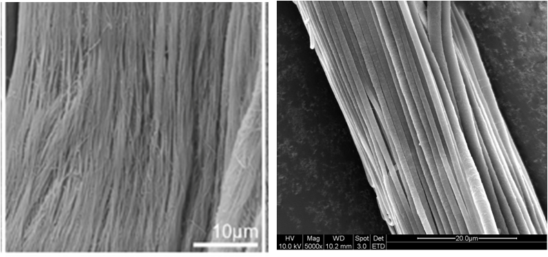
Role of Fiber Alignment

The membranes in the study were described as aligned nanofibers. Fiber alignment can influence cell orientation and migration, particularly in musculoskeletal applications where anisotropic tissue architecture is critical. While the study focuses primarily on hydrophilicity effects, alignment may contribute to guiding tissue organization at the interface.

Considerations for Surface Modification

Beyond polymer blending, electrospinning platforms allow additional strategies such as incorporation of bioactive agents or post-fabrication surface treatments. However, the Ren et al. work specifically highlights that even without complex biochemical functionalization, physicochemical modulation alone can significantly alter immune response and adhesion outcomes.

Applications and Clinical Relevance

Tendon Repair and Adhesion Prevention

Postoperative adhesions remain a major complication in tendon surgery, limiting mobility and functional recovery. In the rat Achilles tendon injury model used by Ren et al., pure PCL membranes were associated with substantial tissue adhesion. In contrast, PCL/PEG membranes reduced adhesion formation, and the highest PEG ratio yielded the most favorable outcome in terms of reduced inflammation and improved tissue separation.

These findings suggest that electrospun implant interface layers may serve as physical and biological barriers that minimize pathological fibrotic bridging while supporting controlled healing.


Broader Implications for Implant–Tissue Integration

Although the study specifically evaluates a tendon model, the underlying principle—modulation of macrophage phenotype through surface hydrophilicity—has broader implications for other soft tissue implant applications. However, extrapolation to orthopaedic hard-tissue implants or cardiovascular devices requires dedicated experimental validation.

The work supports a paradigm in which implant surface engineering prioritizes immune modulation as a primary design objective.

PCL aligned fibers made at 1000 rpm

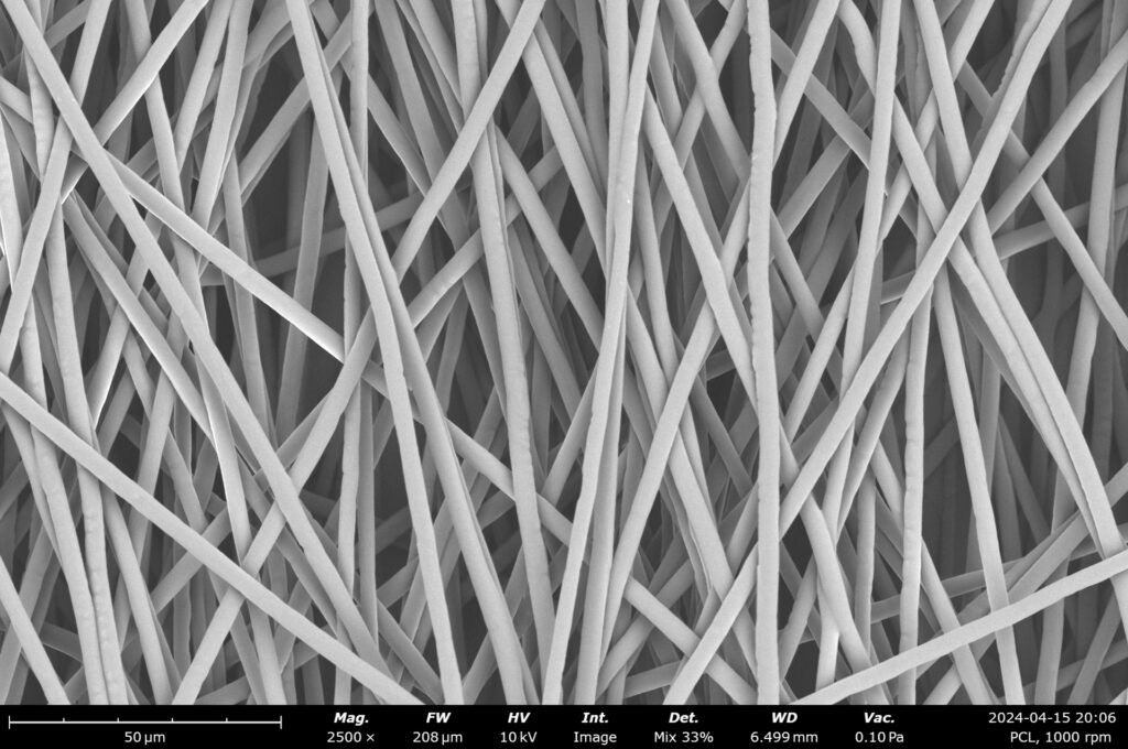
PCL aligned fibers made at 1000 rpm. Image credit: Nanoscience Instruments.

Fluidnatek’s Capabilities in Implant Interface Nanofiber Development

Translating preclinical findings into practical biomedical applications requires reproducible fabrication platforms capable of controlling fiber morphology, alignment, and polymer composition.

Fluidnatek provides electrospinning systems designed to support:

  • Precise control of polymer blending (e.g., PCL/PEG ratios)
  • Fabrication of aligned nanofiber membranes
  • Reproducible control over fiber diameter and morphology
  • Development of degradable fibrous interface layers

Such platforms enable research teams to replicate and extend experimental configurations similar to those described by Ren et al., facilitating systematic studies on implant–tissue integration and immune modulation.

More information on electrospinning platforms for biomedical research is available at: https://fluidnatek.com/electrospinning-machines/

Conclusion: Toward Immunomodulatory Implant Interfaces

Electrospun fibers as implant interface layers represent a strategic evolution in biomedical surface engineering. Rather than functioning solely as passive structural coatings, these nanofiber membranes can actively influence early immune responses and subsequent tissue remodeling.

The study by Ren et al. demonstrates that tuning hydrophilicity through PCL/PEG blending modulates macrophage gene expression and reduces adhesion formation in a rat tendon injury model. Increased PEG content correlated with reduced inflammatory signaling, enhanced M2-like polarization, and fewer postoperative adhesions. Additionally, higher PEG ratios were associated with structural changes consistent with faster degradation, which may facilitate tissue separation at the interface.

These findings reinforce the concept that surface chemistry and nanoscale architecture are central determinants of implant performance. Continued investigation into electrospun nanofiber interface layers may advance the development of next-generation biomedical implants designed not only for mechanical function, but also for precise biological integration.

References

Ren, Y., et al. (2023). Electrospun fibers as implant interface layer. ElectrospinTech. Retrieved from http://electrospintech.com/implantinterface.html

Zhang, X., Liu, L., Wang, Y., & Chen, H. (2021). Electrospun nanofiber scaffolds in regenerative medicine. Acta Biomaterialia, 134, 123–140. https://doi.org/10.1016/j.actbio.2021.04.010

Li, Q., Yang, J., Zhao, Y., & Wang, L. (2020). Electrospun nanofibers as implant coatings for tissue regeneration. Journal of Biomedical Materials Research Part A, 108(9), 1834–1845.


Tissue Engineering: General Introduction to Electrospun Regenerative Scaffolds

Electrospun Regenerative Scaffolds

Introduction: Tissue Regeneration as a Cornerstone of Modern Medicine

Tissue regeneration has become one of the most transformative paradigms in modern medicine, offering a pathway to repair or replace tissues and organs that have been damaged by trauma, degenerative diseases, or surgical interventions. Instead of relying solely on transplants or prosthetic devices, regenerative medicine leverages endogenous healing mechanisms, supported by biomaterials that act as scaffolding designed to facilitate cellular growth and functional tissue integration. Central to this effort is the concept of the electrospun regenerative scaffold—an engineered three-dimensional structure designed to support cellular attachment, migration, proliferation, and differentiation. These scaffolds not only provide physical support but also replicate the biochemical cues of the extracellular matrix (ECM).

Among all available scaffold fabrication technologies, electrospinning has emerged as a frontrunner, enabling the creation of nanofibrous matrices that closely mimic the fibrous architecture of native tissues. The result is a platform with unparalleled control over fiber size, orientation, porosity, and bioactive incorporation.

The electrospun regenerative scaffold represents a fusion of material science, nanotechnology, and biomedical engineering. Its importance continues to grow as researchers and clinicians seek biomimetic, biodegradable, and functional solutions for complex medical needs—from wound care to bone, vascular, and neural regeneration.

What Are Regenerative Scaffolds and Why Electrospinning Excels

A regenerative scaffold can be defined as a supportive matrix that facilitates the growth of new tissue by providing a temporary environment where cells can adhere, proliferate, differentiate, and eventually remodel the matrix into functional native tissue. To ensure functional efficacy, these scaffolds must adhere to rigorous requirements:

  • Biocompatibility to avoid rejection or inflammation.
  • Biodegradability, with degradation rates matching tissue growth.
  • Tunable porosity and fiber architecture to allow cell infiltration and nutrient flow.
  • Mechanical stability to withstand stresses in the target tissue.
  • Bioactivity, achieved by functionalization with peptides, proteins, or growth factors.

Traditional fabrication methods (e.g., freeze-drying, phase separation) can achieve some of these features but often lack precision. Electrospinning, by contrast, allows the production of nanofiber scaffolds with diameters from ~50 nm to 10-20 μm, offering a morphology highly analogous to the ECM.

The advantages of electrospinning for tissue engineering include:

  • Scalability: From lab-scale single-needle systems to industrial multi-jet and free-surface platforms.
  • Material versatility: Natural, synthetic, and hybrid polymers.
  • Customization: Control of fiber alignment, gradient structures, or multi-layer scaffolds.
  • Surface functionalization: Capability to incorporate growth factors, antimicrobials, or nanoparticles.

This versatility ranks electrospun regenerative scaffolds as the most promising platform for next-generation tissue engineering.

Materials and Design Strategies for Electrospun Tissue Scaffolds

Electrospun regenerative scaffolds can be fabricated from a wide range of natural and synthetic polymers, as well as composite blends that optimize specific properties.

  • Natural polymers: Collagen, gelatin, silk fibroin, hyaluronic acid, and chitosan offer intrinsic biocompatibility and promote cell attachment and signaling.
  • Synthetic polymers: Polycaprolactone (PCL), polylactic acid (PLA), poly(lactic-co-glycolic acid) (PLGA), and polyurethane provide predictable mechanical properties and tunable biodegradability.
  • Blended or composite systems: Hybrid scaffolds combine the strengths of both categories. For example, collagen-PCL scaffolds integrate the bioactivity of collagen with the durability of PCL.

Collagen-PCL Nanofibers for Bone or Skin Regeneration

Hybrid collagen-PCL electrospun nanofibers represent one of the most extensively investigated systems.

 Their nanostructure closely mimics native ECM, promoting osteogenic differentiation in bone models or accelerating re-epithelialization in skin regeneration. By adjusting the ratio of collagen to PCL, researchers can fine-tune mechanical strength, porosity, and degradation kinetics can be precisely tailored to meet specific clinical requirements.

Scaffolds for Nerve Guidance and Wound Healing

Aligned electrospun fibers are particularly effective for guiding neurite outgrowth in nerve regeneration. These scaffolds serve as conduits that not only provide physical direction but also deliver biochemical cues. Similarly, electrospun wound healing matrices can incorporate antimicrobial agents, growth factors, or oxygen-releasing nanoparticles to accelerate recovery in complex wounds.

Advanced Design Strategies

Recent innovations include:

  • Core–shell nanofibers for sustained drug release.
  • Macroporous scaffolds achieved by combining electrospinning with 3D printing or salt-leaching.
  • Gradient scaffolds with varying composition or fiber orientation, mimicking tissue interfaces such as tendon-to-bone junctions.

These design strategies push electrospun regenerative scaffolds closer to clinical translation by addressing challenges in cell infiltration, vascularization, and long-term integration.

comparison tendon

Comparison between natural tendon ECM [Youngstrom DW et al 2013] and electrospun nanofibrous bundle showing distinct physical similarity.

Biomedical Applications of Electrospun Scaffolds

Electrospun regenerative scaffolds have shown potential across a wide range of biomedical fields:

  • Bone tissue engineering: Promoting osteoconductivity and vascular ingrowth.
  • Cartilage and tendon repair: Supporting load-bearing structures with aligned nanofibers.
  • Vascular grafts: Providing endothelialization surfaces in small-diameter vessels.
  • Neural repair: Guiding axonal regrowth in peripheral nerve injury.
  • Skin and wound healing: Acting as dressings that prevent infection and stimulate healing.
  • Dental and periodontal regeneration: Serving as bioactive membranes.
  • Cardiac and skeletal muscle regeneration: Mimicking anisotropic fiber orientation for contractile tissues.

Internal link suggestion: See more about Fluidnatek’s regenerative medicine solutions.

External references: Tissue Engineering Part A, Biomaterials, ACS Biomaterials Science & Engineering.

Functionalization Strategies: Beyond Structural Support

While structural biomimicry is essential, advanced regenerative scaffolds also require biofunctionalization to actively influence tissue repair.

Growth Factor Incorporation

Electrospun nanofibers can encapsulate growth factors such as VEGF (vascular endothelial growth factor) or BMP-2 (BMP-2 (bone morphogenetic protein-2), releasing them gradually to stimulate angiogenesis or osteogenesis.

Antimicrobial and Antioxidant Functionalization

In wound healing, scaffolds may integrate silver nanoparticles, copper oxide, or natural antimicrobials to prevent infection. Antioxidants such as curcumin or vitamin E-loaded fibers protect cells from oxidative stress.

Drug-Loaded Electrospun Fibers

Controlled drug delivery through electrospun scaffolds allows localized treatment of infections, cancer, or inflammatory conditions, reducing systemic side effects.

Hybrid Platforms with Biofabrication

Recent approaches combine electrospinning with 3D bioprinting or hydrogel integration, producing hybrid platforms where mechanical support and biological function are seamlessly combined.

From Research to Clinic: The Role of Scalable Electrospinning

One of the greatest challenges in tissue engineering is translation from laboratory-scale proof-of-concept to clinical-grade production. This requires reproducibility, scalability, and regulatory compliance.

Fluidnatek’s electrospinning platforms are designed for this transition:

  • Precise process control for fiber morphology and reproducibility.
  • Multi-material spinning enabling gradient scaffolds and functionalized fibers.
  • Closed systems compliant with GMP (Good Manufacturing Practices).
  • Scalability from R&D to pilot and industrial production.

Beyond equipment, success in clinical translation requires meeting regulatory frameworks:

Internal link suggestion: Discover Fluidnatek’s platforms for clinical scaffold development.

Conclusion

The electrospun regenerative scaffold is reshaping the future of tissue engineering, combining biomimicry, versatility, and scalability. From bone and cartilage repair to neural and vascular regeneration, these scaffolds provide an ECM-like environment that supports cell growth and integration. With advanced functionalization strategies, they extend beyond passive matrices to become bioactive, therapeutic platforms.

As clinical translation accelerates, scalable and regulatory-compliant electrospinning systems such as those developed by Fluidnatek are essential to bring laboratory discoveries into hospitals and patient care.

Looking to develop next-generation regenerative scaffolds? Fluidnatek’s electrospinning platforms empower researchers and biomedical companies to design, functionalize, and scale ECM-like nanofiber scaffolds for advanced clinical applications.

References

  1. Owida HA, Safina R, El-Ghobashy M, Elgendy H. Recent Applications of Electrospun Nanofibrous Scaffold in Biomedical Science. Biomedicines. 2022 Feb;10(2):294.
  2. Han S, Kim J, Park J. 3D Electrospun Nanofiber‐Based Scaffolds: From Fabrication to Applications in Tissue Engineering. Int J Polym Sci. 2021;8790143.
  3. Zhang Y, Zhang M, Cheng D, Xu S, Du C, Xie L, Zhao W. Applications of electrospun scaffolds with enlarged pores in tissue engineering. Biomater Sci. 2022 Mar 15;10(6):1423–1447.
  4. Huang T et al. Application and Development of Electrospun Nanofiber Scaffolds for Bone Tissue Engineering. ACS Biomaterials Sci Eng. 2024 Jun.
  5. Ma Y, Zhang W, Chen G. Electrospinning-based bone tissue scaffold construction. Materials & Design. 2025.
  6. Suamte L et al. Electrospun Based Functional Scaffolds for Biomedical Applications. ScienceDirect. 2024.
  7. Fluidnatek. Electrospun scaffolds for bone tissue engineering. 2024.

For further reading, explore featured articles in Biomaterials and Tissue Engineering Part A.

Case Study — Evonik & VECOLLAN®: Recombinant Collagen Nanofiber Manufacturing Through Electrospinning with Fluidnatek® LE-50

VECOLLAN Fluidnatek

Animal-Free Alternatives in Biomedical Materials

The biomedical sector is undergoing a decisive transition toward fully animal-free materials for regenerative medicine, advanced wound care, and premium cosmetic technologies. This shift is driven not only by ethical considerations but also by growing regulatory requirements for full traceability, pathogen safety, and reproducible manufacturing processes.

In this context, Evonik has developed VECOLLAN®—a recombinant collagen-like peptide designed for biomedical applications. VECOLLAN® is produced through a scalable, reproducible fermentation-based process and offers exceptional purity, safety, and consistency.

In a recent study, Evonik utilized VECOLLAN® to create electrospun meshes using the Fluidnatek® LE-50 equipment—a versatile electrospinning platform for advanced research and pilot-scale process optimization. The LE-50 enabled a coaxial electrospinning setup, placing VECOLLAN® in the fiber core while distributing a controlled crosslinking agent in the outer shell. This configuration delivered three key benefits:

  • Enhanced mechanical stability of the scaffold
  • Reduced swelling in biological environments
  • Tunable dissolution behavior

These properties are critical for implantable devices, controlled drug-release platforms, and next-generation wound care solutions.

This case study demonstrates how Fluidnatek® systems empower the development of next-generation biomaterials—consistent, safe, sustainable. The LE-50’s flexibility, environmental control, and compatibility with post-processing integrations make it an essential tool for organizations seeking to accelerate innovation while minimizing process risk and time to market.

👉 Official Evonik publication: Recombinant collagen platforms 

  1. Krauss C, Montero Mirabet M, Zhang JF, Mader K. Electrospinning of animal-free derived collagen-like protein: Development and characterization of VECOLLAN(R)- nanofibers for biomedical applications. Int J Pharm X. 2025;10:100398.

Electrospun Bioresorbable Tubular Scaffolds for Advanced Medical Devices

electrospun bioresorbable tubular scaffolds

Introduction: The Need for Biofunctional Medical Devices

Electrospinning has emerged as a transformative technology in biomedical engineering, enabling the fabrication of nanofibrous materials that closely mimic the hierarchical structure and functionality of the extracellular matrix (ECM) found in native tissues. This biomimetic capability is particularly valuable for developing next-generation medical devices including vascular grafts, stent coatings, bioresorbable stents, nerve conduits, and electrospun bioresorbable tubular scaffolds. These applications demand precise control over material architecture, mechanical properties, biocompatibility, and degradation kinetics to achieve optimal functional performance.

The growing demand for minimally invasive, patient-specific interventions has accelerated interest in electrospun tubular constructs that can be fully resorbed by the body after fulfilling their therapeutic function. This application note explores the current state of electrospinning technology for producing electrospun bioresorbable tubular scaffolds, highlights key applications in medical device development, and discusses emerging trends in this rapidly evolving field.


Electrospinning Technology for Bioresorbable Tubular Scaffold Production

Process Fundamentals

Electrospinning for tubular scaffold fabrication involves applying a high voltage (10-30 kV) to a polymer solution or melt, creating an electrostatic force that overcomes surface tension to form a jet. This jet undergoes whipping and stretching as the solvent evaporates, resulting in nanofibers that collect on a opposite voltage rotating mandrel to form tubular structures. The process allows precise control over:

  • Fiber diameter (typically 100-500 nm)
  • Fiber orientation (random or aligned)
  • Porosity (60-90%)
  • Wall thickness (50 μm to several mm)
  • Mechanical properties (tensile strength, compliance, and elasticity)
  • Surface chemistry and topography

Equipment Configurations

Several commercial systems have been developed specifically for tubular scaffold production, including the Fluidnatek LE-100 Bio Tubing platform. These advanced electrospinning systems typically feature:

  • Multiple collector options: Rotating mandrels with variable diameters (0.5-10 mm) and rotation speeds (50-2000 rpm) for seamless tubular scaffold fabrication
  • In-line monitoring: Real-time thickness measurement and fiber morphology analysis for stringent quality control
  • Environmental control: Precision regulation of temperature (18-45°C) and humidity (10-80% RH) to ensure reproducibility
  • Clean processing environments: ISO 5/Class 100 compatible chambers for aseptic, contamination-free processing
  • Automation capabilities: Programmable deposition patterns and process parameters for complex architectures
  • Data management: Industry 4.0 integration for process traceability and validation

Materials for Electrospun Bioresorbable Scaffolds

The selection of appropriate polymers is critical for successful bioresorbable scaffold development. Commonly used materials include:

Polymer

Degradation Time

Key Properties

Common Applications

Poly(lactic acid) (PLA)

12-24 months

High strength, moderate hydrophobicity

Vascular grafts, bone scaffolds

Poly(glycolic acid) (PGA)

2-4 months

Rapid degradation, good cell adhesion

Nerve guides, temporary stents

Poly(lactic-co-glycolic acid) (PLGA)

1-12 months (tunable)

Controllable degradation rate

Drug delivery, soft tissue engineering

Polycaprolactone (PCL)

24-36 months

Excellent elasticity, slow degradation

Long-term vascular applications

Polyurethanes (PU)

Variable

Superior mechanical properties

Heart valves, vascular devices

Natural polymers (collagen, silk, chitosan)

Variable

Enhanced bioactivity

Tissue engineering, wound healing

Multi-material approaches using polymer blends or core-shell configurations enable tailored degradation profiles and mechanical properties specific to each application.

Electrospun Scaffolds for Medical Devices and Tissue Engineering

Electrospun bioresorbable tubular scaffolds are advancing several areas in medical device development:

Electrospun Vascular Grafts

Electrospun vascular grafts represent a promising alternative to autologous vessels for bypass procedures and vascular repair. Their advantages include:

  • Tunable compliance: Matching mechanical properties with native vessels reduces hemodynamic disturbances and intimal hyperplasia
  • Controlled porosity: Optimized pore size (typically 10-30 μm) facilitates cell infiltration while maintaining barrier function
  • Drug delivery capabilities: Incorporation of anticoagulants, anti-inflammatories, or growth factors enhances performance
  • Degradation synchronized with tissue regeneration: Scaffold provides initial support and gradually transfers load to newly formed tissue

Clinical studies have demonstrated promising results for small-diameter (<6 mm) vascular grafts, with ongoing trials for peripheral and coronary applications.

Stent Coatings and Fully Bioresorbable Stents

Electrospun polymeric coatings for conventional metal stents (including nitinol-based stents) as well as fully bioresorbable stent platforms offer several advantages:

  • Controlled drug elution: Precise release kinetics for antiproliferative agents
  • Reduced foreign body response: Gradual dissolution minimizes chronic inflammation
  • Preservation of vessel geometry: After resorption, native vessel mechanics are restored
  • Facilitation of repeat interventions: Absence of permanent implants simplifies future procedures
  • Enhanced compatibility with nitinol stents: Electrospun coatings can mitigate nickel ion release while maintaining the mechanical advantages of nitinol.

Recent innovations include dual-layer electrospun stents with different drug release profiles and mechanical properties in each layer[8].

Nerve Conduits and Neural Tissue Engineering

Tubular electrospun conduits support nerve regeneration following injury by:

  • Directing axonal growth: Aligned nanofibers guide regenerating neurons
  • Preventing scar tissue infiltration: Semipermeable walls block fibroblast migration
  • Supporting Schwann cell migration: Optimized architecture promotes cellular colonization
  • Delivering neurotrophic factors: Sustained release of growth factors enhances nerve regeneration

Electrospun nerve guides have shown promising results in peripheral nerve defects up to 30 mm in preclinical models.

Hybrid Metal-Polymer Scaffolds

An important advancement in electrospun scaffold technology is the development of hybrid constructs combining metallic frameworks with electrospun polymer coatings. Nitinol (nickel-titanium alloy) is particularly valuable in these applications due to its unique properties:

  • Shape memory effect: Allows for minimally invasive deployment and self-expansion
  • Superelasticity: Provides mechanical support while maintaining flexibility
  • Biocompatibility: Well-established safety profile in vascular applications
  • Fatigue resistance: Withstands physiological cyclic loading

Electrospun coatings on nitinol structures can:

  • Deliver therapeutic agents locally
  • Modulate the tissue-material interface
  • Provide a template for tissue ingrowth
  • Create a barrier to control nitinol ion release

These hybrid constructs are particularly valuable for stents, occlusion devices, and embolic protection systems where the mechanical properties of nitinol complement the biological functionality of electrospun polymers[10].

Other Emerging Applications

Additional applications leveraging electrospun bioresorbable tubular scaffolds include:

  • Tracheal and bronchial replacement: Reinforced electrospun constructs with radial rigidity and longitudinal flexibility
  • Gastrointestinal stents: Degradable stents for temporary stricture management
  • Urethral reconstruction: Tailored scaffolds supporting regeneration of functional urethral tissue
  • Drug delivery conduits: Tubular implants for localized, sustained therapeutic delivery

Manufacturing Considerations

Quality Control Parameters

Consistent performance of electrospun tubular scaffolds depends on rigorous quality control focused on:

  • Structural uniformity: Even fiber distribution and orientation throughout the scaffold
  • Mechanical consistency: Batch-to-batch reproducibility of tensile strength, burst pressure, and compliance
  • Chemical purity: Residual solvent levels below regulatory thresholds (<50 ppm for common solvents)
  • Sterility assurance: Validated sterilization processes compatible with delicate nanostructures

Scale-Up Strategies

Transitioning from laboratory to commercial production requires addressing several challenges:

  • Throughput enhancement: Multinozzle or needleless systems to increase production volume
  • Process validation: Design of Experiments (DoE) approaches to establish robust process parameters
  • Inline monitoring: Real-time quality verification systems for continuous production
  • Regulatory compliance: Documentation systems meeting cGMP, ISO 13485, and FDA requirements
  • Sterilization compatibility: Process development for terminal sterilization methods preserving scaffold integrity

Regulatory Considerations

Electrospun bioresorbable scaffolds face specific regulatory challenges:

  • Novel material combinations: May require additional biocompatibility and degradation testing
  • Long-term degradation products: Assessment of tissue response to breakdown components
  • Process validation: Critical process parameters for electrospinning must be thoroughly documented
  • Mechanical testing standards: Often requires development of custom test methods specific to the intended application
  • Shelf-life determination: Stability of both mechanical properties and biological activity must be demonstrated

Regulatory pathways differ by region and specific application, with combination products (incorporating drugs or biologics) facing more complex requirements.

Clinical Case Studies

Case Study 1: Small-Diameter Vascular Grafts

A recent clinical trial evaluated PCL/PLA electrospun grafts (4 mm diameter) for hemodialysis access in 12 patients. Key findings included:

  • 83% primary patency at 6 months
  • No aneurysm formation or catastrophic mechanical failure
  • Progressive endothelialization observed via ultrasound
  • Degradation profile matching tissue ingrowth rates

Case Study 2: Drug-Eluting Bioresorbable Stent Coating

A PLGA electrospun coating on a metal stent platform demonstrated:

  • Reduced restenosis rates compared to bare metal stents (8% vs. 22%)
  • Complete resorption by 9 months post-implantation
  • Reduced dual antiplatelet therapy requirements
  • Improved vessel healing and reduced inflammation

Future Trends and Challenges

Several emerging approaches are poised to advance electrospun tubular scaffold technology:

  • Smart responsive scaffolds: Integration of stimuli-responsive materials that adapt to physiological changes
  • 4D printing approaches: Electrospun structures programmed to change shape or properties over time
  • Hybrid manufacturing: Combining electrospinning with other fabrication techniques (3D printing, textile processes)
  • Cell electrospinning: Direct incorporation of living cells during the fabrication process
  • Personalized medicine applications: Patient-specific scaffold designs based on medical imaging data

Challenges requiring further research include:

  • Mechanical property optimization: Matching complex native tissue mechanics more precisely
  • Control of degradation heterogeneity: Ensuring uniform resorption throughout the scaffold volume
  • Scale-up limitations: Addressing throughput constraints for high-volume applications
  • Standardization: Developing consensus testing methods specific to electrospun materials


Conclusion

Electrospun bioresorbable tubular scaffolds represent a significant advancement in medical device technology, offering unprecedented control over scaffold architecture, material properties, and biological response. As manufacturing capabilities continue to mature and clinical evidence accumulates, these materials are positioned to address critical unmet needs in vascular, neural, and other tubular tissue applications. Continued innovation in materials, processing techniques, and hybrid approaches will further expand the potential of this versatile technology platform.

Designed for Excellence in Tubular Scaffold Manufacturing
The Fluidnatek LE-100 BioTubing system is specially engineered to meet the stringent requirements of tubular scaffold production. Its advanced rotating mandrel system, precision-controlled environment, and high-resolution deposition capabilities enable the fabrication of seamless, uniform, and reproducible tubular structures. With full GMP-compliant architecture and options for cleanroom integration, the LE-100 BioTubing is the ideal platform for scaling up from research to clinical manufacturing of bioresorbable vascular grafts, nerve conduits, and other implantable devices.

Let’s Build the Future of Medical Devices
Are you developing resorbable scaffolds for advanced biomedical applications

**Fluidnatek’s electrospinning platforms** deliver the precision, reproducibility, and scalability needed to design **customised tubular nanostructures** for next-generation medical devices. 

👉 Contact our team (https://fluidnatek.com/contact) to discuss your biomedical project.

References

  1. Zhang Y, et al. Recent advances in electrospinning for biomedical applications. Biomater Sci. 2022;10(2):316-339. https://doi.org/10.1039/D1BM01518C
  2. Sensini A, et al. Hierarchical electrospun tendon-ligament bioinspired scaffolds. Biofabrication. 2023;15(1):015004. https://doi.org/10.1088/1758-5090/aca8c6
  3. Keirouz A, et al. Nanofiber-based wound dressings and their applications. Mater Sci Eng C. 2023;113:111018. https://doi.org/10.1016/j.msec.2020.111018
  4. Khorshidi S, et al. A review of key challenges of electrospun scaffolds for tissue-engineering applications. J Tissue Eng Regen Med. 2022;16(3):195-215. https://doi.org/10.1002/term.3267
  5. Gao S, et al. Core-shell nanofibers: Nano channel and capsule by coaxial electrospinning. Adv Mater Interfaces. 2023;10(7):2101770. https://doi.org/10.1002/admi.202101770
  6. Nagarajan S, et al. Design strategies for controlling degradation rate and mechanical properties in electrospun vascular scaffolds. ACS Appl Mater Interfaces. 2022;14(41):45829-45843. https://doi.org/10.1021/acsami.2c09274
  7. Fukunishi T, et al. Tissue-engineered small diameter arterial vascular grafts from cell-free nanofiber PCL/chitosan scaffolds in a sheep model. PLoS One. 2022;17(3):e0254315. https://doi.org/10.1371/journal.pone.0254315
  8. Qiu X, et al. Controlled dual-drug release from electrospun nanofibers as bioresorbable local drug delivery systems. J Control Release. 2023;353:607-618. https://doi.org/10.1016/j.jconrel.2022.12.039
  9. Wang S, et al. Aligned electrospun polycaprolactone/silk fibroin core-shell nanofibers for nerve tissue engineering. J Biomed Mater Res A. 2023;111(5):814-826. https://doi.org/10.1002/jbm.a.37487
  10. Torres-Giner S, et al. Industrial applications of electrospinning: Drug delivery, tissue engineering, and regulatory considerations. Int J Mol Sci. 2023;24(4):3676. https://doi.org/10.3390/ijms24043676
  11. Tsetsekou M, et al. Nitinol-polymer composites for medical applications: A review. J Mater Sci. 2023;58(10):4692-4721. https://doi.org/10.1007/s10853-022-08128-1
  12. Kuznetsov K, et al. Surface modification of nitinol stents with electrospun bioresorbable polymers: Approaches and clinical outcomes. J Biomater Appl. 2022;37(3):481-496. https://doi.org/10.1177/08853282221131975

Electrospun Wound Dressing: A Breakthrough in Advanced Wound Healing

wound-dressing-electrospinning

Electrospinning has emerged as a transformative technology for designing next-generation wound dressings. The unique ability of this technique to produce nanofiber-based scaffolds that mimic the extracellular matrix (ECM) has positioned it at the forefront of biomedical research. As chronic wounds, burns, and post-surgical injuries demand increasingly sophisticated care, electrospun wound dressings offer unmatched potential for accelerating healing, preventing infections, and delivering therapeutic agents in a controlled manner.

The Clinical Challenge in Wound Care

Chronic and acute wounds remain a significant clinical burden, particularly among aging populations and individuals with diabetes, vascular disease, or immunocompromised states. Conventional dressings often fail to provide optimal moisture retention, mechanical protection, or antimicrobial activity. Furthermore, they rarely support cellular activities required for tissue regeneration.

In contrast, nanofiber wound dressing systems can be engineered to address these limitations through structural mimicry of native tissue, functional loading with bioactive compounds, and controlled drug release. The growing body of research and innovation in biomedical electrospinning highlights the urgent need for advanced wound dressing materials.

human skin wound

View of a human skin wound.

Benefits of Electrospun Nanofibers for Wound Care

Electrospinning enables the production of continuous fibers with diameters ranging from tens of nanometers to a few micrometers, offering several biomedical advantages:

Mimicking the Extracellular Matrix (ECM)

The fibrous architecture of electrospun mats closely resembles the ECM, providing a favorable environment for cell adhesion, proliferation, and differentiation. This promotes effective re-epithelialization and granulation tissue formation.

Tunable Porosity and Moisture Control

By adjusting parameters such as voltage, flow rate, and polymer concentration, the porosity of the electrospun membrane can be finely tuned. This facilitates gas exchange while preventing bacterial infiltration, which is vital for wound healing.

Functionalization with Bioactive Agents

Nanofiber scaffolds can be functionalized with antimicrobial agents, growth factors, and anti-inflammatory drugs, enabling drug-loaded electrospun fibers that actively participate in the healing process rather than serving as passive barriers.

Mechanical Adaptability

Electrospun mats can be designed with elasticity and strength suitable for various anatomical sites, from joints to pressure points, enhancing patient comfort and compliance.


Polymeric Systems and Functionalization Strategies

The choice of polymers significantly influences the properties and functionality of electrospun wound dressings. Both synthetic and natural polymers are employed, often in blends to balance biocompatibility, degradability, and mechanical performance.

Synthetic Polymers for Structural Integrity

Polymers such as polycaprolactone (PCL), poly(lactic acid) (PLA), and polyurethane (PU) are frequently used due to their mechanical robustness and processability. These materials ensure the scaffold maintains structural integrity over time.

Biopolymers for Antimicrobial Effect and Bioactivity

Natural polymers, including collagen, gelatin, chitosan, and hyaluronic acid, offer inherent bioactivity. Biopolymer wound dressing systems leverage these materials to introduce antimicrobial and hemostatic properties.

For instance, chitosan is widely recognized for its antimicrobial properties and has been incorporated into nanofibrous matrices to enhance wound healing efficacy PubMed source.


Drug Delivery and Bioactive Capabilities

Electrospinning facilitates controlled drug release by embedding pharmaceuticals within or on the surface of the nanofibers. This delivery mode ensures a sustained release at the wound site, improving therapeutic outcomes and reducing systemic side effects.

Release Kinetics and Porosity Design

By modulating the polymer composition and fiber morphology, researchers can customize release profiles ranging from burst release to prolonged delivery over several days or weeks. Porosity design plays a critical role in mediating this process and can be optimized for different wound types and stages.

Multi-drug and Layered Systems

Advanced configurations such as core–shell nanofibers, multilayered mats, and coaxial spinning enable incorporation of multiple drugs with staggered release kinetics. This is especially valuable in treating infected wounds or those requiring both antimicrobial and regenerative agents.

Examples include loading electrospun mats with silver nanoparticles for antibacterial effects alongside vascular endothelial growth factor (VEGF) for tissue regeneration ScienceDirect source.

Vascular endothelial growth factor A (VEGF A) protein molecule

Vascular endothelial growth factor A (VEGF A) protein molecule. Cartoon representation combined with semi transparent surfaces.

Clinical Potential and Future Perspectives

The translation of electrospinning for biomedical applications from bench to bedside is accelerating. Several preclinical studies and early-stage clinical trials highlight the promising outcomes of wound healing scaffolds based on electrospun materials.

Regulatory Considerations

Despite the promise, regulatory hurdles persist. Sterilization techniques, reproducibility of fiber architecture, and scalability for mass production are key challenges. However, platforms like Fluidnatek® electrospinning systems are designed to meet Good Manufacturing Practice (GMP) requirements, easing the path to commercialization.

Personalized and Smart Dressings

Future directions point toward personalized wound care solutions, integrating biosensors for real-time monitoring, stimuli-responsive drug release, and AI-assisted design of scaffold parameters based on wound morphology.

Innovative research in wound healing biomaterials is increasingly leveraging machine learning and big data analytics to fine-tune material properties for individualized therapy.


Conclusion: From Research to Clinical Application

Electrospun wound dressings are reshaping the landscape of wound management. Their unique combination of biomimetic structure, bioactivity, and versatility makes them ideal candidates for a wide range of clinical applications—from diabetic ulcers to battlefield injuries.

As the field progresses, the synergy between material science, bioengineering, and medical practice will drive the development of even more effective solutions.

Are you exploring advanced wound care materials? Discover how Fluidnatek’s electrospinning platforms help design, test and scale biomedical nanofiber dressings tailored to your research or product needs. Explore our biomedical electrospinning solutions.


References

  1. Chouhan, D., & Mandal, B. B. Silk biomaterials in wound healing and skin regeneration therapeutics: From bench to bedside. Acta Biomaterialia, 2020, 103, 24–51. DOI: 10.1016/j.actbio.2019.11.050
  2. Boateng, J. S., Matthews, K. H., Stevens, H. N. E., & Eccleston, G. M. Wound healing dressings and drug delivery systems: A review. Journal of Pharmaceutical Sciences, 2008, 97(8), 2892–2923. DOI: 10.1002/jps.21210
  3. Zhang, Y. Z., Venugopal, J., Huang, Z. M., Lim, C. T., & Ramakrishna, S. Crosslinking of the electrospun gelatin nanofibers. Polymer, 2006, 47(8), 2911–2917. DOI: 10.1016/j.polymer.2006.02.046
  4. Li, X., Kanjwal, M. A., Lin, L., & Chronakis, I. S. Electrospun polyvinyl-alcohol nanofibers as oral fast-dissolving delivery system of caffeine and riboflavin. Colloids and Surfaces B: Biointerfaces, 2013, 103, 182–188. DOI: 10.1016/j.colsurfb.2012.10.023
  5. Zhang, H., He, P., Kang, Y., & Wang, L. Electrospun composite nanofibers for functional wound dressings: A review. Journal of Industrial Textiles, 2022, 52(2), 1–30. DOI: 10.1177/15280837221106633
  6. Chen, S., Li, R., Li, X., Xie, J. Electrospinning: An enabling nanotechnology platform for drug delivery and regenerative medicine. Advanced Drug Delivery Reviews, 2018, 132, 188–213. DOI: 10.1016/j.addr.2018.07.002
  7. Khorshidi, S., Karkhaneh, A., A review on nanofiber scaffolds for wound healing applications. Journal of Biomedical Materials Research Part A, 2018, 106(9), 2530–2545. DOI: 10.1002/jbm.a.36483
  8. Yarin, A. L. Coaxial electrospinning and emulsion electrospinning of core–shell fibers. Polymer, 2011, 52(9), 2029–2044. DOI: 10.1016/j.polymer.2011.02.042

Cancer Detection and Diagnosis Using Electrospun Fibers

Cancer detection electrospun fibers

The early detection and accurate diagnosis of cancer remain critical challenges in modern healthcare. Despite technological advances, many cancers are still diagnosed at late stages, compromising treatment effectiveness and patient survival rates. But electrospun fibers have a lot to say on this subject.

Among the innovative technologies being developed, electrospun fibers have emerged as revolutionary materials for creating highly sensitive biosensors and diagnostic platforms.

This article explores how electrospun nanofibers are transforming cancer detection through enhanced sensitivity, specificity, and rapid response times.

Electrospun Fibers: What They Are and How They Work

Electrospun fibers are ultrafine filaments produced through a versatile technique called electrospinning, which utilizes electrical forces to draw charged threads from polymer solutions or melts. The resulting fibers typically have diameters ranging from nanometers to micrometers, creating materials with exceptional characteristics due to their resemblance to human tissues, ideal for biomedical applications, particularly cancer biosensing.

The electrospinning process involves:

  1. A polymer solution loaded into a syringe with a metal needle
  2. One or more high-voltage power supplies (typically 5-30 kV)
  3. A grounded or negatively charged collector plate or rotating mandrel
  4. Precise environmental control (temperature, humidity)

When voltage is applied, the polymer solution becomes charged, and when electrostatic repulsion overcomes surface tension, a jet erupts from the needle tip. As this jet travels toward the collector, the solvent evaporates, leaving behind solid polymer fibers that form a non-woven mesh or membrane.

These electrospun nanofibers exhibit several key properties that make them exceptional for cancer detection:

  • Extremely high surface-to-volume ratio, enhancing biomarker capture efficiency
  • Tunable porosity for controlled molecular interactions
  • Customizable fiber diameter and orientation
  • Ability to incorporate functional materials (antibodies, enzymes, nanoparticles)
  • Three-dimensional architecture that mimics the extracellular matrix (ECM)

Fluidnatek’s electrospinning technology enables precise adjustment of fiber diameter, porosity, and surface chemistry—attributes crucial for creating effective biosensors that are sensitive, cost-effective, and suitable for point-of-care testing.

Applications of Electrospun Fibers in Cancer Detection

The versatility of electrospun fibers has enabled their integration into multiple cancer detection platforms. These applications leverage the unique structural and functional properties of nanofibers to identify cancer biomarkers with unprecedented sensitivity.

Some of these applications include:

Electrospun Nanofiber Scaffolds for Cancer Cell Detection

Early detection of cancer cells can dramatically improve patient outcomes. Traditional diagnostic methods often lack the sensitivity to detect low-abundance biomarkers in bodily fluids. Electrospun nanofibers address this limitation by providing:

  • A three-dimensional architecture that mimics the extracellular matrix (ECM), supporting cell adhesion and growth
  • The ability to be functionalized with biomolecular probes (such as antibodies or aptamers) for high selectivity toward cancer-specific markers

For instance, studies have demonstrated that nanofiber membranes functionalized with prostate-specific membrane antigen (PSMA)-targeted ligands can selectively capture prostate cancer cells from mixed populations. These captured cells can then be analyzed using fluorescence imaging or molecular assays, resulting in improved detection speed and accuracy compared to conventional methods.

Cancer_detection

Fluorescence pictures of cancer biomarkers on electrospun PS substrates obtained by an inverted fluorescence microscope (200×). (A) AFP (DyLight 488, green), (B) CEA (DyLight 405, blue), (C) VEGF (DyLight 649, red); (a-c) light field, (d-f) fluorescence field, (g-i) superposition view of the two fields. Wang et al (2013) PLoS ONE 2013; 8(12): e82888.

Functionalization Strategies for Selective Detection

Functionalizing electrospun membranes is essential for selective cancer cell detection. Several techniques have proven effective:

  • Surface Chemistry Engineering: Methods such as plasma treatment, chemical grafting, and layer-by-layer deposition provide precise control over surface properties. For instance, membranes modified with antibodies against PSMA show high specificity for prostate cancer cells.
  • Multiplexed Detection: Advanced approaches integrate multiple biomarkers onto a single electrospun membrane, enabling simultaneous detection of various cancer types. This multiplexing is particularly valuable when cancer markers overlap across different tumor types, enhancing diagnostic accuracy.

Integration into Microfluidic Systems

Combining electrospun nanofibers with microfluidic chips allows for the development of compact diagnostic devices capable of real-time cancer monitoring. These lab-on-a-chip systems integrate sample processing, detection, and data analysis, making them ideal for point-of-care applications in clinical settings or resource-limited environments.

Case Studies and Recent Advances

Circulating Tumor Cell Capture Using Electrospun Platforms

CTCs, (Circulating tumor cells) are cancer cells that detach from primary tumors and enter the bloodstream, playing a critical role in the metastatic spread of cancer. Their detection and isolation offer valuable insights for early diagnosis, prognosis, and personalized treatment strategies. Electrospun fiber meshes, particularly when functionalized with tumor-specific antibodies (such as anti-EpCAM), have demonstrated remarkable efficiency in capturing these rare cells directly from blood samples.

The unique architecture of electrospun nanofibers—featuring high surface-area-to-volume ratios, tunable porosity, and a 3D interconnected structure—creates an optimal microenvironment for cell capture. These characteristics enable greater interaction between the fibers and flowing blood, increasing the likelihood of CTC adhesion. Recent studies have shown that well-engineered electrospun platforms can achieve capture rates exceeding 90%, significantly outperforming conventional flat-surface or microfluidic-based systems. In one of them, published by Lab on a Chip by Chen, L., et al. (2017), the researchers developed a microfluidic device integrated with electrospun poly (lactic-co-glycolic acid) (PLGA) nanofibers functionalized with anti-EpCAM antibodies.

The high surface area and 3D structure of the nanofibers significantly enhanced the contact between the target cells and the capture surface. The platform achieved capture efficiencies above 90% for EpCAM-positive CTCs in spiked blood samples. The system also maintained high viability of captured cells, enabling downstream analysis.

Functionalization plays a key role in the capture mechanism: antibodies or aptamers immobilized on the nanofiber surfaces selectively bind to antigens expressed on CTC membranes. As blood flows through or across the fibrous mat, CTCs are selectively retained, while most normal blood cells pass through. This specificity and efficiency make electrospun platforms highly promising for liquid biopsy applications and real-time cancer monitoring.

Applications in Liquid Biopsy

Liquid biopsy, a minimally invasive technique analyzing biomarkers from blood, is transforming cancer diagnostics. Electrospun fibers enhance this approach by serving as solid-phase platforms to capture rare cancer cells or exosomes from complex fluids.

A groundbreaking study published in PLoS ONE by Wang et al. (2013) demonstrated the use of electrospun polystyrene (PS) substrates for detecting multiple cancer biomarkers simultaneously. The researchers successfully detected alpha-fetoprotein (AFP), carcinoembryonic antigen (CEA), and vascular endothelial growth factor (VEGF) using fluorescence microscopy on functionalized nanofiber scaffolds, showing the potential for multiplexed cancer detection on a single platform.

Multi-Biomarker Detection Systems

Recent advances in electrospinning for cancer detection have led to the development of systems capable of detecting multiple biomarkers simultaneously. For example, researchers have created electrospun polyacrylonitrile (PAN) fibers functionalized with different antibodies that can detect breast cancer markers like HER2, ER, and PR from a single sample, enabling more accurate subtyping of breast cancers.

Smart Responsive Nanofibers

“Smart” responsive materials have been incorporated into electrospun nanofibers to create visual detection systems. A notable example is the development of pH-responsive polymeric nanofibers that change color in the presence of metabolic byproducts from cancer cells, enabling naked-eye detection without sophisticated equipment.

Advantages of Electrospun Fibers Over Other Cancer Detection Technologies

We must emphasize that electrospun nanofibers offer several significant advantages over conventional cancer detection technologies:

Enhanced Sensitivity and Lower Detection Limits

The high surface-to-volume ratio of electrospun fibers dramatically increases the density of biorecognition elements, improving sensitivity. Comparative studies show that electrospun membranes outperform traditional diagnostic materials such as flat films or hydrogels in several ways:

  • Faster cell capture kinetics
  • Improved detection limits (down to sub-nanomolar concentrations)
  • Lower sample volume requirements
  • Enhanced mechanical stability for repeated use

Improved Specificity Through Surface Modification

The surface of electrospun nanofibers can be easily modified with multiple recognition elements (antibodies, aptamers, molecularly imprinted polymers) to enhance specificity and reduce false positives. This multi-recognition approach has been particularly effective in distinguishing between closely related cancer subtypes.

Point-of-Care Applicability

Unlike many conventional cancer detection systems that require specialized laboratory equipment, electrospun fiber-based biosensors can be designed for point-of-care use. Their flexible, portable nature makes them suitable for use in clinics, remote areas, or even home-based monitoring systems.

Cost-Effectiveness and Scalability

Clearly, the electrospinning process is relatively simple and cost-effective compared to other nanofabrication techniques. The equipment required is less expensive than that needed for techniques like photolithography or electron beam lithography, making electrospun nanofiber technologies more accessible for widespread implementation in cancer diagnostics.

External Validation and Scientific Support

A review published in ACS Applied Materials & Interfaces2 confirms that nanofiber-based platforms enhance biosensing sensitivity by closely mimicking biological microenvironments. This external validation supports the growing adoption of electrospun fibers for next-generation cancer diagnostics.

Challenges and Future Directions in Electrospun Biosensors

Despite promising progress, several challenges must be addressed to translate electrospun fiber biosensors from laboratory research to clinical practice:

  • Scalability: Ensuring reproducibility across production batches
  • Regulatory compliance: Thorough assessment of biocompatibility and toxicity
  • Long-term stability: Maintaining membrane sensitivity over extended periods

Current research in electrospinning biomedical applications is focused on:

  1. Smart polymers that respond to specific biomolecular interactions
  2. Real-time readout electronics for continuous monitoring
  3. AI-based data analysis to improve diagnostic accuracy
  4. Biodegradable nanofibrous scaffolds for in vivo cancer sensing
  5. Multi-functional nanofibers that combine detection with therapeutic agent delivery

As these technologies mature, we can expect increasingly sensitive, specific, and user-friendly cancer diagnostic tools based on electrospun nanofibers.

Conclusion: The Future of Cancer Detection Using Electrospun Fibers

Electrospun fibers represent a revolutionary approach to cancer detection and diagnosis, offering unprecedented sensitivity, specificity, and versatility. Their unique structural properties and adaptability make them ideal platforms for developing next-generation cancer biosensors.

As research advances and clinical validation progresses, these electrospun nanofibers will likely play an increasingly important role in early cancer detection efforts, potentially transforming patient outcomes through earlier intervention.

The continued development of electrospinning for cancer detection exemplifies how advanced materials science can address critical healthcare challenges, bridging the gap between laboratory innovation and clinical application. By enabling earlier and more accurate diagnoses—potentially even before symptoms arise—electrospun membranes are poised to become a cornerstone in personalized cancer diagnostics.

If your research team is exploring electrospun nanofibers for biosensor development or cancer diagnostic applications, contact Fluidnatek to learn how our advanced electrospinning technologies can support your research and scale-up efforts. Our precision platforms empower researchers to develop tailored solutions for complex biomedical challenges, from proof-of-concept to commercial scalability.

References

  1. Zhang N, Deng Y, Tai Q, et al. (2012). Electrospun TiO2 Nanofiber-Based Cell Capture Assay for Detecting Circulating Tumor Cells from Colorectal and Gastric Cancer Patients. Advanced Materials. 24(20):2756-2760. https://pubmed.ncbi.nlm.nih.gov/22528884/
  2. Wang X, Wang G, Liu G, et al. (2002). Electrospun Nanofibrous Membranes for Highly Sensitive Optical Sensors. ACS Applied Materials & Interfaces. 8(41):28150-28155. DOI: 10.1021/acsami.6b10269 https://pubs.acs.org/doi/10.1021/nl020216u
  3. Huang, Z-M., Zhang, Y-Z., Kotaki, M., & Ramakrishna, S. (2003). A review on polymer nanofibers by electrospinning and their applications in nanocomposites. Composites Science and Technology, 63(15), 2223–2253. https://doi.org/10.1016/S0266-3538(03)00178-7
  4. Noh, H., Lee, S. H., & Kim, J. (2020). Recent advances in nanofiber-based biosensors for biomedical applications. Biosensors and Bioelectronics, 148, 111800. https://doi.org/10.1016/j.bios.2019.111800
  5. Liu, Y., et al. (2020). Electrospun nanofibers for sensors and wearable electronics: a review. Materials Today, 41, 168–193. https://doi.org/10.1016/j.mattod.2020.08.005
  6. Jiang, Y., et al. (2017). Electrospun nanofiber membranes for efficient cancer cell capture. ACS Applied Materials & Interfaces, 9(12), 11350–11358. https://doi.org/10.1021/acsami.6b15025
  7. ElectrospinTech. (n.d.). Electrospun Membranes for Cancer Cell Detection. Recuperado de: http://electrospintech.com/cancerdetect.html
  8. Wang, L., et al. (2021). Functional electrospun nanofibers for cancer diagnostics. Advanced Functional Materials, 31(20), 2100212. https://doi.org/10.1002/adfm.202100212
  9. Fluidnatek. (2024). Applications of Electrospinning in Biomedical Engineering. https://www.fluidnatek.com/applications

Electrospun Scaffolds for Bone Treatment and Repair: A Breakthrough in Bone Tissue Engineering

Electrospun Scaffolds for Bone Tissue

Electrospun scaffolds for bone tissue engineering have emerged as a groundbreaking solution for treating and repairing bone defects. This innovative approach combines advanced materials science with bioengineering principles to create scaffolds that mimic the natural extracellular matrix (ECM) of bone tissue, promoting regeneration and healing

What is Electrospinning and How Does It Work?

Firstly, Electrospinning is a versatile technique that uses electrical forces to produce fine fibers from polymer solutions or melts. The process involves applying a high voltage to a polymer solution made of a polymer and at least one solvent, which is then drawn into ultrafine fibers due to electrical repulsion as it travels towards a grounded collector. This method allows for precise control over fiber diameter, orientation, and composition, making it ideal for creating scaffolds that closely resemble the structure of natural bone tissue.

Applications of Electrospun Fibers in Bone Tissue Engineering

Electrospun scaffolds for bone tissue engineering

For instance, Electrospun scaffolds provide an ideal environment for bone cell growth and differentiation. These scaffolds offer high surface-area-to-volume ratios, porosity, and compositional diversity, which are essential for mimicking the extracellular matrix of natural bone. Recent advancements have addressed challenges such as cell infiltration and 3D tissue formation through innovative techniques like sharp inclined array collectors with point electrodes.

Electrospun bio-nanocomposite scaffolds for bone tissue engineering

Identically, bio-nanocomposite scaffolds combine synthetic or natural polymers with bioactive inorganic materials to enhance mechanical strength and osteoconductivity. For example, incorporating hydroxyapatite nanoparticles into PVA/PVP scaffolds improves cell adhesion and calcium deposition. Additionally, zirconium-reinforced composites have shown increased compressive strength while maintaining cytocompatibility.

Electrospun submicron bioactive glass fibers for bone tissue scaffold

Nonetheless, bioactive glass fibers have gained attention for their ability to bond with bone and stimulate angiogenesis. These fibers, composed of silicon dioxide, calcium oxide, and phosphorus pentoxide, release ions crucial for bone formation. Studies have shown that bioactive glass-PCL composites demonstrate significantly higher alkaline phosphatase activity compared to polymer-only scaffolds, indicating accelerated mineralization.

Electrospun scaffolds preparation

Electrospun scaffolds for bone tissue engineering have emerged as a groundbreaking solution for treating and repairing bone defects. This innovative approach, particularly, combines advanced materials science with bioengineering principles to create scaffolds that mimic the natural extracellular matrix (ECM) of bone tissue, promoting regeneration and healing.

Advantages of Using Electrospun Fibers to Repair Bone

Certainly, Electrospun nanofibers for bone regeneration offer several advantages over traditional bone repair methods:

  1. Biomimetic structure: Electrospun fibers closely mimic the natural extracellular matrix of bone tissue, providing an ideal environment for cell growth and differentiation.
  2. Tailored properties: The electrospinning process allows for precise control over fiber diameter, orientation, and composition, enabling the creation of scaffolds with optimized mechanical and biological properties.
  3. Enhanced cell adhesion and proliferation: The high surface-area-to-volume ratio of electrospun scaffolds promotes cell attachment and growth.
  4. Controlled drug delivery: Electrospun fibers can be loaded with growth factors, antibiotics, or other therapeutic agents for sustained release, enhancing bone regeneration and reducing infection risks. This approach offers several advantages:
    1. Localized delivery: Moreover, the scaffolds can provide targeted release of drugs directly to the bone defect site, maximizing therapeutic efficacy.
    2. Sustained release profiles: By carefully selecting polymer-drug combinations and fiber architectures, release kinetics can be tailored to match the healing process, from initial inflammation to long-term bone remodeling.
    3. Multi-drug delivery: Different drugs can be incorporated into various fiber populations or layers within the scaffold, allowing for sequential or simultaneous release of multiple therapeutic agents.
    4. Protection of sensitive biomolecules: The fibrous structure can shield growth factors and other delicate compounds from degradation, preserving their bioactivity.
    5. Reduced systemic side effects: Localized, controlled release minimizes the need for high systemic drug doses, potentially decreasing adverse effects.
    6. Infection control: Antibiotics can be incorporated to create an antimicrobial environment, crucial for preventing post-operative infections in bone repair procedures.
    7. Synergistic effects: The combination of scaffold architecture and drug delivery can work synergistically to promote cell infiltration, vascularization, and ultimately, bone regeneration
  5. Customizable degradation rates: By selecting appropriate materials and repair processing parameters, the degradation rate of electrospun scaffolds can be tailored to match the rate of new bone formation.

Future Perspectives in Bone Tissue Regeneration

Specifically, the future of electrospun scaffolds for bone tissue engineering looks promising, with several emerging trends:

  1. Multifluid electrospinning: Advanced techniques like coaxial and triaxial systems enable the creation of layered fiber architectures with spatially controlled bioactive agents.
  2. 4D dynamic scaffolds: Temperature and pH-responsive fibers that can adapt their pore size post-implantation to accommodate tissue ingrowth are being developed.
  3. AI-driven fabrication: Researchers are employing machine learning algorithms to optimize process parameters and predict scaffold morphology and mechanical performance.
  4. Integration with other technologies: Combining electrospinning with 3D printing, melt electrowriting, electrospraying, and microfluidics is opening new possibilities for creating complex, multifunctional scaffolds.

Overall, the combination of electrospinning and 3D printing or melt electrowriting leverages the strengths of both techniques:

  1. Enhanced structural complexity: 3D printing provides precise control over macrostructure, while electrospinning adds nanofiber layers that mimic the extracellular matrix.
  2. Improved mechanical properties: The integration results in scaffolds with both adequate mechanical strength from 3D-printed structures and high porosity from electrospun fibers.
  3. Hierarchical architectures: This approach allows for the creation of scaffolds with multi-scale features, from nanometer to millimeter ranges.
  4. Fabrication methods:
    • Direct electrospinning onto 3D-printed structures
    • Alternating layers of 3D-printed and electrospun materials
    • Using electrospun nanofibers as a component in 3D printing inks

Conclusion

After all, as research in this field continues to advance, electrospun scaffolds for bone tissue engineering are poised to revolutionize bone treatment and repair, offering personalized solutions for complex bone defects and bridging the gap between laboratory research and clinical application.

In order to learn more about the latest developments in electrospun nanofibers for bone regeneration, check out this comprehensive review from ACS Biomaterials Science & Engineering.

Interested in how electrospinning technology can advance bone tissue engineering? Contact us to explore tailored solutions.

References

  1. Bhardwaj, N., & Kundu, S. C. (2010). Electrospinning: A fascinating fiber fabrication technique. Biotechnology Advances, 28(3), 325-347.
  2. Khajavi, R., Abbasipour, M., & Bahador, A. (2016). Electrospun biodegradable nanofibers scaffolds for bone tissue engineering. Journal of Applied Polymer Science, 133(3), 42883.
  3. Langer, R., & Vacanti, J. P. (1993). Tissue engineering. Science, 260(5110), 920-926.
  4. Li, W. J., Laurencin, C. T., Caterson, E. J., Tuan, R. S., & Ko, F. K. (2002). Electrospun nanofibrous structure: A novel scaffold for tissue engineering. Journal of Biomedical Materials Research, 60(4), 613-621.
  5. Pham, Q. P., Sharma, U., & Mikos, A. G. (2006). Electrospinning of polymeric nanofibers for tissue engineering applications: A review. Tissue Engineering, 12(5), 1197-1211.
  6. Sill, T. J., & von Recum, H. A. (2008). Electrospinning: Applications in drug delivery and tissue engineering. Biomaterials, 29(13), 1989-2006.
  7. Teo, W. E., & Ramakrishna, S. (2006). A review on electrospinning design and nanofibre assemblies. Nanotechnology, 17(14), R89-R106.
  8. Zafar, M., Najeeb, S., Khurshid, Z., Vazirzadeh, M., Zohaib, S., Najeeb, B., & Sefat, F. (2016). Potential of electrospun nanofibers for biomedical and dental applications. Materials, 9(2), 73.

Cell-Seeded Scaffolds: Revolutionizing Biomedical Engineering for Tissue Regeneration

Cell-Seeded Scaffold

For decades, researchers in biomedical engineering have strived to unlock the secrets of tissue engineering and tissue regeneration. The ultimate goal: to repair or replace damaged tissues and organs, offering hope to millions suffering from injuries and diseases. One of the most promising approaches in this field involves the creation of cell-seeded scaffolds, structures that mimic the natural environment of cells and guide their growth and development.

Cell-Seeded Scaffolds

The Promise of Cell-Seeded Scaffolds

Imagine a tiny, three-dimensional framework, meticulously designed to support the growth of new tissue. That’s essentially what a cell-seeded scaffold is. These scaffolds provide structural support for cells to attach, proliferate, and differentiate, ultimately forming functional tissue. The beauty of this approach lies in its potential to create personalized, biocompatible implants that seamlessly integrate with the body.

But how are these scaffolds made, and what makes them so effective? The answer lies in a combination of advanced materials science, cell biology, and innovative fabrication techniques.

Electrospinning: A Key Technology for Scaffold Fabrication

Among the various methods used to create tissue affolds, electrospinning stands out as a versatile and powerful technique. This process uses an electrical field to draw charged threads of polymer solutions, creating nanofibers that form a porous, three-dimensional structure. The resulting scaffolds closely resemble the extracellular matrix (ECM), the natural environment surrounding cells in the body.

Advantages of Electrospinning in Tissue Engineering

The advantages of electrospinning for biomedical tissue engineering are numerous:

  • Tunable architecture: Electrospinning allows for precise control over fiber diameter, porosity, and alignment, enabling the creation of scaffolds tailored to specific tissue types.
  • Versatile materials: A wide range of polymers, both natural and synthetic, can be electrospun, allowing for the selection of materials with specific properties such as biodegradability, biocompatibility, and mechanical strength.

  • Scalability: The electrospinning process can be scaled up for mass production, making it a viable option for clinical applications.

Collagen Electrospinning: A Natural Choice

Collagen, the most abundant protein in the human body, is a popular choice for electrospinning scaffolds. Its inherent biocompatibility, biodegradability, and ability to promote cell adhesion make it an ideal material for tissue engineering applications. Collagen electrospinning cell seeding techniques are thus widely studied.

Applications of Collagen Scaffolds

Collagen scaffolds can be used to regenerate and repair different tissues, including:

  • Skin: Collagen scaffolds can promote wound healing and reduce scarring.
  • Bone: They can guide the formation of new bone tissue for fracture repair and bone regeneration.
  • Cartilage: They can support the growth of chondrocytes (cartilage-forming cells) for treating osteoarthritis and other cartilage defects.
  • Blood vessels: Collagen scaffolds can be used to create vascular grafts for bypass surgery and other cardiovascular applications.

Bio-Electrospinning: Seeding Cells During Scaffold Formation

While traditional methods involve seeding cells onto pre-fabricated scaffolds, a more advanced approach—known as bio-electrospinning cell seeding—integrates cells directly into the electrospinning process. This technique involves suspending cells in the polymer solution and simultaneously electrospinning the polymer while encapsulating the cells within the fibers.

Benefits of Bio-Electrospinning

The benefits of bio-electrospinning are significant:

  • Enhanced cell viability: Encapsulating cells within fibers protects them from harsh conditions during electrospinning, improving their survival rate.
  • Uniform cell distribution: Bio-electrospinning ensures homogenous distribution of cells throughout the scaffold, promoting uniform tissue formation.
  • Improved cell-matrix interactions: Direct encapsulation allows intimate contact between cells and scaffold material, enhancing adhesion, proliferation, and differentiation.
Characterization of the 3DPCL-GelMA Scaffold

Characterization of the 3DPCL-GelMA Scaffold. (a) A scanning electron microscope (SEM) image showing the cross-section of melt electrospun polycaprolactone (PCL) fibers, arranged in a porous network. The scale bar represents 30 µm. (b) An SEM image depicting a 3D-printed PCL-GelMA (PG) scaffold composed of 50 stacked layers, highlighting its organized microstructure. The scale bar represents 200 µm. (e) A 3DPCL-GelMA scaffold with cells co-cultured, illustrating cell attachment and distribution within the scaffold structure. (f) A 3DPCL-GelMA scaffold after the hydrogel component has been removed, revealing the remaining fibrous architecture. Reference: Kong et al., 2024.

Beyond the Basics: Advanced Electrospinning Techniques

Researchers are continuously developing new electrospinning techniques to further enhance scaffold properties. Some advanced approaches include:

Coaxial Electrospinning

This technique uses two concentric needles to create core-shell fibers. It allows encapsulation of cells or growth factors within the core fiber structure for controlled release or targeted delivery.

Melt Electrospinning Writing (MEW)

MEW offers precise control over molten polymer deposition. This enables highly defined 3D scaffolds with controlled architecture and mechanical properties.

Combining Electrospinning with Electrospraying

Combining electrospinning with electrospraying produces 3D scaffolds that incorporate stem cells directly into their structure. This technique enhances cell integration within scaffolds.

Hybrid 3D Printing and Electrospinning

This method combines 3D printing with electrospinning to fabricate complex tissue structures like vascular patches or organ-like constructs.

These advanced techniques offer unprecedented control over scaffold properties and cell behavior, paving the way for more effective therapies in biomedical engineering tissue regeneration.

histological cross-sections of scaffolds seeded with cells

Illustration of histological cross-sections of scaffolds seeded with cells after different cultivation periods: (A) After 1 day, showing initial cell attachment and distribution (magnification: ×200). (B) After 15 days, revealing enhanced cell proliferation and scaffold integration (magnification: ×100). Scale bars: 50 µm. [Braghirolli et al., 2015].

Challenges and Future Directions

While cell-seeded scaffolds hold immense promise for tissue engineering, several challenges remain:

  • Scalability: Scaling up production while maintaining quality is critical for clinical translation.
  • Vascularization: Engineering functional blood vessels within scaffolds is essential for nutrient delivery.
  • Immune response: Minimizing immune reactions is vital for long-term success.

Future Research Goals

Future research efforts will focus on:

  • Developing biomaterials with improved biocompatibility.
  • Incorporating bioactive molecules like growth factors into scaffolds.
  • Designing more sophisticated scaffold architectures that mimic native tissues.
  • Promoting vascularization strategies while minimizing immune responses.

Conclusion

Cell-seeded scaffolds represent a groundbreaking advancement in biomedical engineering. By combining innovative technologies like collagen electrospinning cell seeding with advanced fabrication techniques such as bio-electrospinning or coaxial electrospinning, researchers are pushing the boundaries of what’s possible in regenerative medicine. With continued innovation, these technologies could revolutionize treatments for injuries and diseases—bringing us closer to a future where personalized tissue implants are readily available.

References:

Author: Wee-Eong TEO

  1. Ang H Y, Irvine S A, Avrahami R, Sarig U, Bronshtein T, Zussman E, Boey F Y C, Machluf M, Venkatraman. Characterization of a bioactive fiber scaffold with entrapped HUVECs in coaxial electrospun core-shell fiber. Biomatter 2014; 4: e28238. View
  2. Braghirolli D I, Zamboni F, Acasigua G A X, Pranke P. Association of electrospinning with electrospraying: a strategy to produce 3D scaffolds with incorporated stem cells for use in tissue engineering. International Journal of Nanomedicine 2015; 10: 5159. 
  3. Erben J, Jirkovec R, Kalous T, Klicova M, Chvojka J. The Combination of Hydrogels with 3D Fibrous Scaffolds Based on Electrospinning and Meltblown Technology. Bioengineering. 2022; 9(11):660. 
  4. Kong X, Zhu D, Hu Y, Liu C, Zhang Y, Wu Y, Tan J, Luo Y, Chen J, Xu T, Zhu L. Melt electrowriting (MEW)-PCL composite Three-Dimensional exosome hydrogel scaffold for wound healing. Materials & Design 2024; 238: 112717. 
  5. Lee H, Kim G H. Enhanced cellular activities of polycaprolactone/alginate-based cell-laden hierarchical scaffolds for hard tissue engineering applications. Journal of Colloid and Interface Science 2014; 430: 315.

Visionary solutions: electrospun implants giving new hope to nerve recovery

Implantes Electrospun en la Recuperación de Nervios Periféricos

The Role of Biomaterials in Treating Peripheral Nerve Injury

Peripheral nerve injury (PNI) remains a significant medical challenge due to its slow recovery process and complex clinical outcomes. When a nerve is damaged, prolonged denervation can lead to muscle atrophy and reduced Schwann cell activity, both critical for axonal regeneration. In response, innovative approaches such as biomaterial-based implants have emerged as promising solutions to accelerate nerve recovery.

While drugs like ibuprofen have shown potential in promoting nerve regeneration through anti-inflammatory properties, systemic administration often causes unwanted side effects. To overcome this, electrospinning in the biomedical field has gained traction as a method for delivering drugs directly to the injury site via polymer-based scaffolds. Recently, the University College London School of Pharmacy published a study in which the team developed ibuprofen-loaded electrospun materials suitable for surgical implantation in peripheral nerve injuries using our Fluidnatek LE-50 G2 equipment.

What is Electrospinning and Why is it Ideal for Nerve Recovery?

Electrospinning is a versatile technique that transforms polymer solutions into fine, nano- to micro-scale fibers by applying a high-voltage electric field. These fibers are collected into mats that mimic the extracellular matrix of tissues, making them ideal candidates for biomedical applications, especially in nerve repair.

The advantages of electrospun materials include:

  1. Customizability: Physical properties like mechanical strength and drug release rates can be tuned.
  2. Biocompatibility: Synthetic polymers such as polycaprolactone (PCL) and polylactic acid (PLA) are widely used due to their compatibility with biological systems.
  3. Sustained Drug Release: Electrospun fibers can encapsulate drugs like ibuprofen, ensuring controlled and prolonged release at the target site.

For peripheral nerve injury, electrospun wraps or implants loaded with therapeutic agents significantly enhance the healing process by delivering localized treatment, minimizing side effects.

Electrospinning and Ibuprofen Delivery for Nerve Recovery

Recent advancements have demonstrated the successful development of ibuprofen-loaded electrospun biomaterials for peripheral nerve injury. Ibuprofen, a widely used non-steroidal anti-inflammatory drug (NSAID), is known to improve nerve regeneration by inhibiting inflammatory responses and promoting neurite growth.

In a cutting-edge study, researchers optimized the use of electrospun nerve wraps fabricated from PCL, PLA, and their copolymers. The following findings underscore the potential of these polymer-based implants:

  • Optimized Fiber Properties: Electrospinning parameters were tuned to produce smooth, defect-free fibers with varying diameters. The incorporation of ibuprofen into these fibers allowed for a controlled, sustained release over 21 days.
  • Surgical Handling: User evaluations highlighted the importance of mechanical properties, with PLA/PCL (70/30) blends demonstrating superior flexibility and strength, making them ideal for nerve-wrapping applications.
  • In Vivo Performance: In animal models, ibuprofen-loaded electrospun materials accelerated nerve regeneration. Axon counts in treated nerves were significantly higher compared to controls, confirming the therapeutic effect of localized ibuprofen delivery.
electrospun material implantation procedure in a rat sciatic nerve crush model.

Photographs showing stages of electrospun material implantation procedure in a rat sciatic nerve crush model.

Polymer Selection in Electrospinning for Biomedical Implants

The success of electrospun biomaterials depends heavily on the choice of polymers. For peripheral nerve injury, polymers must exhibit biocompatibility, biodegradability, and mechanical stability. The following polymers are commonly employed:

  1. Polylactic Acid (PLA): Known for its slow degradation rate, PLA provides a robust structure but can be brittle.
  2. Polycaprolactone (PCL): Offers excellent flexibility and strength, ideal for implants requiring pliability.
  3. PLA/PCL Copolymers: Combining the strengths of PLA and PCL, these copolymers achieve the desired balance of mechanical stability and handling ease.

In the case of ibuprofen-loaded electrospun implants, PLA/PCL (70/30) was identified as the most suitable formulation due to its superior surgical handling and sustained drug release profile.

Summary of formulation properties

Summary of formulation properties. Scanning electron micrographs (A) reveal cylindrical fibres with no visible defects. A histogram of fibre diameters (B) shows unimodal distribution for all tested formulations. Cumulative ibuprofen release data (C) present an initial burst release followed by a period of sustained release over 21 days (Each formulation was tested in triplicate, and the results are presented as mean ± SEM (n = 3)).

The Future of Electrospun Biomaterials in Nerve Repair

As research in the biomedical field advances, electrospinning continues to demonstrate immense potential for improving outcomes in nerve injuries. Key areas of future development include:

  • Scalable Manufacturing: Ensuring that electrospun materials can be mass-produced for clinical use.
  • Advanced Drug Loading: Incorporating multiple therapeutic agents for synergistic effects on nerve regeneration.
  • Clinical Trials: Translating promising in vivo results into human applications to validate the efficacy and safety of electrospun biomaterials.

Conclusion

The use of electrospinning in the biomedical field has revolutionized the development of drug-loaded implants for peripheral nerve injury. By leveraging polymers such as PLA and PCL, researchers have created biomaterials capable of delivering sustained, localized treatment, accelerating nerve regeneration and functional recovery.

Ibuprofen-loaded electrospun fibers represent a significant advancement in nerve recovery strategies, offering a targeted, effective, and minimally invasive solution. As the field continues to evolve, these innovative biomaterials hold the promise of transforming peripheral nerve injury treatment and enhancing patient outcomes.

References

Karolina Dziemidowicz, Simon C. Kellaway, Owein Guillemot-Legris, Omar Matar, Rita Pereira Trindade, Victoria H. Roberton, Melissa L.D. Rayner, Gareth R. Williams, James B. Phillips,

Development of ibuprofen-loaded electrospun materials suitable for surgical implantation in peripheral nerve injury,

Biomaterials Advances,

Volume 154, 2023, 213623,

ISSN 2772-9508,

*All images in the article are the property of the authors.

DRUG-LOADED ELECTROSPUN YARNS OF APPLICATION AS ANTIMICROBIAL SURGICAL SUTURES

Electrospun surgical sutures

Electrospinning for Drug-Loaded Surgical Sutures

The objective of the research work presented in this application note is to develop a suture thread composed of fibers obtained by electrospinning (surgical suture yarns made of electrospun fibers) and interwoven using an electrospun fiber-yarn collector. Ciprofloxacin has been added as an antimicrobial agent to prevent surgical site infections.

In other words, electrospinning technology is positioned as a high-potential alternative for the development of surgical sutures constructed from nanofibers (electrospun nanofibers in this case).

The polymer chosen for the yarn is PHBV, with three variants containing different 3HV unit contents tested.

A complete characterization of the different suture yarns (i.e., the different electrospun yarns obtained) has been carried out, evaluating both their mechanical properties and antimicrobial efficacy. The results show promising mechanical properties and a high antimicrobial effect.

Antimicrobial Properties of Ciprofloxacin-Loaded Electrospun Yarns

Sutures are a routine surgical procedure used to close wounds and join tissues. Due to their intrinsic characteristics, these interventions are susceptible to the emergence of pathogens, leading to what are known as surgical site infections (SSIs).

SSIs cause a large number of medical complications, as well as increased morbidity, mortality, and associated healthcare costs. Over time, a variety of suture threads have been developed based on the characteristics of the tissue to be sutured.

However, despite the significant drawbacks of SSIs, little progress has been made in improving the therapeutic effect of suture threads to prevent these infections.

Generally, the incorporation of antimicrobial substances has been carried out using techniques such as melt spinning, dip coating, or soaking, among others. While these techniques have been shown to be effective to some extent, none of them properly encapsulate the substance of interest. As a result, the release profile and stability over time are not adequately controlled.

This is where the electrospinning technique can provide additional value in the development of drug-loaded electrospun yarns.

Electrospinning for Drug Delivery: A Novel Approach to Encapsulating Bioactive Compounds

In this sense, the technique that has proven to be able to incorporate drugs effectively is electrospinning. Electrospinning allows obtaining nanostructures and microstructures that can incorporate drugs into their polymeric matrix in a single step, thus substantially improving their release process. In addition, another great advantage of using electrospinning for this application is that it is not necessary to use high temperatures to obtain nanofibers, which makes it possible to encapsulate compounds such as proteins, growth factors, peptides, DNA or other substances that would not be possible to encapsulate with other techniques such as melt spinning.

In this scientific contribution, members of Bioinicia‘s R&D department develop a suture thread composed of nanofibers obtained by electrospinning and interwoven by means of a device called electrospun fiber-yarn collection module, an accessory developed by Fluidnatek (Bioinicia Fluidnatek being a subsidiary of the Bioinicia Group), to which ciprofloxacin has been added as an antimicrobial agent.

Mechanical Performance of Electrospun Suture Yarns

There are different biopolymers used in biomedical applications. From PLLA, considered the gold standard, to PEG, PLGA, PDS, PLA or PHA. All of them are polymers that can be processed by electrospinning. Within the PHAs family, which is a biodegradable and highly biocompatible polymer, much research has been done on PHB and, within this, on its copolymer PHBV (poly(3-hydroxybutyrate-co-3-hydroxyvalerate). PHB has high crystallinity and macromolecular organization, resulting in a rigid and brittle material that lacks mechanical strength. On the other hand, PHBV co-polyester shows improved thermal and mechanical properties, which vary depending on the content of 3HV units present in the polyester.

In this work, ciprofloxacin hydrochloride (CPX) has been used as an antimicrobial substance to encapsulate electrospun nanofibers. CPX is an antibiotic belonging to the fluoroquinolone family, with known efficacy against Gram-positive and Gram-negative bacteria.

In this study, 3 types of suture threads based on PHBV polymer with different contents of 3HV units, namely 2%, 10% and 20% molar, have been developed. In all cases, PHBV has been dissolved at 8% wt in TFE (2,2,2-trifluoroethanol). CPX was added at 20% wt in the ratio to the amount of polymer.

Electrospinning for Suture Yarn Production: Process, Equipment, and Material Analysis

The throughput production pilot plant can also implement the electrospun fiber-yarn collector module in its configuration. The solution, contained in a syringe, is pushed by a pump until it emerges from the needle tip.

The high electric field present between the needle tip and the collector elongates the solution, forming a jet due to the electric field’s action. This increases the contact surface between the solution and the medium, causing the evaporation of the solvent and the creation of nanofibers. This is the fundamental process behind the electrospinning technique.

To generate the suture yarns, an accessory called the fiber yarn collector module, developed by Fluidnatek, has been used. This accessory consists of a rotating funnel over which the nanofibers generated by electrospinning are directed.

As the nanofibers approach the funnel, they intertwine following the direction of rotation, eventually forming a yarn that is continuously collected by a rotating reel.

To ensure consistency and reproducibility in the manufacturing of the suture yarns, an Environmental Control Unit (ECU) has been used. The ECU sets specific temperature and relative humidity values—30°C and 30% RH, respectively.

The Environmental Control Unit has also been developed by Fluidnatek, specifically designed for its electrospinning equipment and the unique evaporative process of electrospinning (and electrospraying).

This version makes the information easier to follow while keeping technical details intact.

The mechanical and antimicrobial properties of the different drug-loaded yarns made of electrospun fibers obtained have been evaluated by SEM (scanning electron microscopy) imaging, Fourier transform infrared spectroscopy, wide-angle X-ray scattering, differential scanning calorimetry and in vitro drug release monitoring.

Results and conclusion

The 3 suture yarns generated by electrospinning from PHBV with different concentrations of 3HV units and loaded with CPX show a cylindrical morphology with a total diameter between 300 and 500 μm, composed in turn of individual fibers obtained by the electrospinning process, each of these fibers in turn with an average diameter between 1 and 3 μm. CPX appears in an amorphous state within the yarns and the crystallinity of the polymer decreases as the content of 3HV units increases, which in turn is related to the drug release profile. The presence of CPX in the threads has shown high antibacterial activity for two typical pathogens, one Gram-positive and the other Gram-negative, so these suture threads could be suitable in surgical procedures to prevent SSIs.

Despite the promising mechanical properties and the high antimicrobial effect, the elasticity of the suture yarns generated so far does not reach that of traditional suture yarns, so this parameter should be improved in the future so that this type of yarn could be an alternative to those currently used. But what is clear is that electrospinning is positioned as a serious alternative to produce continuous fiber yarns, and in the specific case that applies to this application note to produce drug-loaded electrospun fiber yarns for medical purposes.

References

Pharmaceutics 2024, 16(2), 220

https://doi.org/10.3390/pharmaceutics16020220

INTERESTED? CONTACT OUR SPECIALISTS!