


Introduction: Tissue Regeneration as a Cornerstone of Modern Medicine
Tissue regeneration has become one of the most transformative paradigms in modern medicine, offering a pathway to repair or replace tissues and organs that have been damaged by trauma, degenerative diseases, or surgical interventions. Instead of relying solely on transplants or prosthetic devices, regenerative medicine leverages endogenous healing mechanisms, supported by biomaterials that act as scaffolding designed to facilitate cellular growth and functional tissue integration. Central to this effort is the concept of the electrospun regenerative scaffold—an engineered three-dimensional structure designed to support cellular attachment, migration, proliferation, and differentiation. These scaffolds not only provide physical support but also replicate the biochemical cues of the extracellular matrix (ECM).
Among all available scaffold fabrication technologies, electrospinning has emerged as a frontrunner, enabling the creation of nanofibrous matrices that closely mimic the fibrous architecture of native tissues. The result is a platform with unparalleled control over fiber size, orientation, porosity, and bioactive incorporation.
The electrospun regenerative scaffold represents a fusion of material science, nanotechnology, and biomedical engineering. Its importance continues to grow as researchers and clinicians seek biomimetic, biodegradable, and functional solutions for complex medical needs—from wound care to bone, vascular, and neural regeneration.
What Are Regenerative Scaffolds and Why Electrospinning Excels
A regenerative scaffold can be defined as a supportive matrix that facilitates the growth of new tissue by providing a temporary environment where cells can adhere, proliferate, differentiate, and eventually remodel the matrix into functional native tissue. To ensure functional efficacy, these scaffolds must adhere to rigorous requirements:
- Biocompatibility to avoid rejection or inflammation.
- Biodegradability, with degradation rates matching tissue growth.
- Tunable porosity and fiber architecture to allow cell infiltration and nutrient flow.
- Mechanical stability to withstand stresses in the target tissue.
- Bioactivity, achieved by functionalization with peptides, proteins, or growth factors.
Traditional fabrication methods (e.g., freeze-drying, phase separation) can achieve some of these features but often lack precision. Electrospinning, by contrast, allows the production of nanofiber scaffolds with diameters from ~50 nm to 10-20 μm, offering a morphology highly analogous to the ECM.
The advantages of electrospinning for tissue engineering include:
- Scalability: From lab-scale single-needle systems to industrial multi-jet and free-surface platforms.
- Material versatility: Natural, synthetic, and hybrid polymers.
- Customization: Control of fiber alignment, gradient structures, or multi-layer scaffolds.
- Surface functionalization: Capability to incorporate growth factors, antimicrobials, or nanoparticles.
This versatility ranks electrospun regenerative scaffolds as the most promising platform for next-generation tissue engineering.
Materials and Design Strategies for Electrospun Tissue Scaffolds
Electrospun regenerative scaffolds can be fabricated from a wide range of natural and synthetic polymers, as well as composite blends that optimize specific properties.
- Natural polymers: Collagen, gelatin, silk fibroin, hyaluronic acid, and chitosan offer intrinsic biocompatibility and promote cell attachment and signaling.
- Synthetic polymers: Polycaprolactone (PCL), polylactic acid (PLA), poly(lactic-co-glycolic acid) (PLGA), and polyurethane provide predictable mechanical properties and tunable biodegradability.
- Blended or composite systems: Hybrid scaffolds combine the strengths of both categories. For example, collagen-PCL scaffolds integrate the bioactivity of collagen with the durability of PCL.
Collagen-PCL Nanofibers for Bone or Skin Regeneration
Hybrid collagen-PCL electrospun nanofibers represent one of the most extensively investigated systems.
Their nanostructure closely mimics native ECM, promoting osteogenic differentiation in bone models or accelerating re-epithelialization in skin regeneration. By adjusting the ratio of collagen to PCL, researchers can fine-tune mechanical strength, porosity, and degradation kinetics can be precisely tailored to meet specific clinical requirements.
Scaffolds for Nerve Guidance and Wound Healing
Aligned electrospun fibers are particularly effective for guiding neurite outgrowth in nerve regeneration. These scaffolds serve as conduits that not only provide physical direction but also deliver biochemical cues. Similarly, electrospun wound healing matrices can incorporate antimicrobial agents, growth factors, or oxygen-releasing nanoparticles to accelerate recovery in complex wounds.
Advanced Design Strategies
Recent innovations include:
- Core–shell nanofibers for sustained drug release.
- Macroporous scaffolds achieved by combining electrospinning with 3D printing or salt-leaching.
- Gradient scaffolds with varying composition or fiber orientation, mimicking tissue interfaces such as tendon-to-bone junctions.
These design strategies push electrospun regenerative scaffolds closer to clinical translation by addressing challenges in cell infiltration, vascularization, and long-term integration.
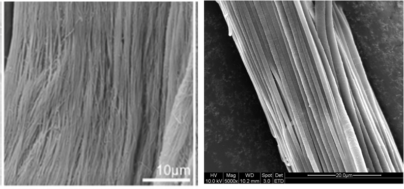
Comparison between natural tendon ECM [Youngstrom DW et al 2013] and electrospun nanofibrous bundle showing distinct physical similarity.
Biomedical Applications of Electrospun Scaffolds
Electrospun regenerative scaffolds have shown potential across a wide range of biomedical fields:
- Bone tissue engineering: Promoting osteoconductivity and vascular ingrowth.
- Cartilage and tendon repair: Supporting load-bearing structures with aligned nanofibers.
- Vascular grafts: Providing endothelialization surfaces in small-diameter vessels.
- Neural repair: Guiding axonal regrowth in peripheral nerve injury.
- Skin and wound healing: Acting as dressings that prevent infection and stimulate healing.
- Dental and periodontal regeneration: Serving as bioactive membranes.
- Cardiac and skeletal muscle regeneration: Mimicking anisotropic fiber orientation for contractile tissues.
Internal link suggestion: See more about Fluidnatek’s regenerative medicine solutions.
External references: Tissue Engineering Part A, Biomaterials, ACS Biomaterials Science & Engineering.
Functionalization Strategies: Beyond Structural Support
While structural biomimicry is essential, advanced regenerative scaffolds also require biofunctionalization to actively influence tissue repair.
Growth Factor Incorporation
Electrospun nanofibers can encapsulate growth factors such as VEGF (vascular endothelial growth factor) or BMP-2 (BMP-2 (bone morphogenetic protein-2), releasing them gradually to stimulate angiogenesis or osteogenesis.
Antimicrobial and Antioxidant Functionalization
In wound healing, scaffolds may integrate silver nanoparticles, copper oxide, or natural antimicrobials to prevent infection. Antioxidants such as curcumin or vitamin E-loaded fibers protect cells from oxidative stress.
Drug-Loaded Electrospun Fibers
Controlled drug delivery through electrospun scaffolds allows localized treatment of infections, cancer, or inflammatory conditions, reducing systemic side effects.
Hybrid Platforms with Biofabrication
Recent approaches combine electrospinning with 3D bioprinting or hydrogel integration, producing hybrid platforms where mechanical support and biological function are seamlessly combined.
From Research to Clinic: The Role of Scalable Electrospinning
One of the greatest challenges in tissue engineering is translation from laboratory-scale proof-of-concept to clinical-grade production. This requires reproducibility, scalability, and regulatory compliance.
Fluidnatek’s electrospinning platforms are designed for this transition:
- Precise process control for fiber morphology and reproducibility.
- Multi-material spinning enabling gradient scaffolds and functionalized fibers.
- Closed systems compliant with GMP (Good Manufacturing Practices).
- Scalability from R&D to pilot and industrial production.
Beyond equipment, success in clinical translation requires meeting regulatory frameworks:
- ISO 10993 for biocompatibility testing.
- FDA and EMA guidelines for medical devices and combination products.
- Sterility and endotoxin testing for implantable scaffolds.
Internal link suggestion: Discover Fluidnatek’s platforms for clinical scaffold development.
Conclusion
The electrospun regenerative scaffold is reshaping the future of tissue engineering, combining biomimicry, versatility, and scalability. From bone and cartilage repair to neural and vascular regeneration, these scaffolds provide an ECM-like environment that supports cell growth and integration. With advanced functionalization strategies, they extend beyond passive matrices to become bioactive, therapeutic platforms.
As clinical translation accelerates, scalable and regulatory-compliant electrospinning systems such as those developed by Fluidnatek are essential to bring laboratory discoveries into hospitals and patient care.
Looking to develop next-generation regenerative scaffolds? Fluidnatek’s electrospinning platforms empower researchers and biomedical companies to design, functionalize, and scale ECM-like nanofiber scaffolds for advanced clinical applications.
References
- Owida HA, Safina R, El-Ghobashy M, Elgendy H. Recent Applications of Electrospun Nanofibrous Scaffold in Biomedical Science. Biomedicines. 2022 Feb;10(2):294.
- Han S, Kim J, Park J. 3D Electrospun Nanofiber‐Based Scaffolds: From Fabrication to Applications in Tissue Engineering. Int J Polym Sci. 2021;8790143.
- Zhang Y, Zhang M, Cheng D, Xu S, Du C, Xie L, Zhao W. Applications of electrospun scaffolds with enlarged pores in tissue engineering. Biomater Sci. 2022 Mar 15;10(6):1423–1447.
- Huang T et al. Application and Development of Electrospun Nanofiber Scaffolds for Bone Tissue Engineering. ACS Biomaterials Sci Eng. 2024 Jun.
- Ma Y, Zhang W, Chen G. Electrospinning-based bone tissue scaffold construction. Materials & Design. 2025.
- Suamte L et al. Electrospun Based Functional Scaffolds for Biomedical Applications. ScienceDirect. 2024.
- Fluidnatek. Electrospun scaffolds for bone tissue engineering. 2024.
For further reading, explore featured articles in Biomaterials and Tissue Engineering Part A.


 ESP
ESP